Iron Toxicity Post #42: K.I.S.S. Keep it simple searchers!

Iron Toxicity Post #42: K.I.S.S. Keep it simple searchers!

One of our clients, Twila J Starkey, and I were chatting the other day about the many posts on this iron subject. 

Her scintillating observation: “There’s just too much there! ”

And I agree.

This post is intended to Keep It Simple, Searchers (K.I.S.S.).

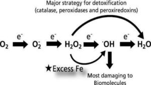
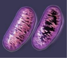
The pic above presents a compelling and straightforward diagram of what excess; unbound iron does inside our body, our organs, our tissue, and ultimately, our cells. So what happens is iron, when not properly bound by transport or regulatory proteins, is pro-oxidant, and thus, very toxic to our health.

Yes, I’ve said that before.

The pic below is intended to show just how ridiculously complex the process of oxidative stress can be. It comes from this epic penetrating article by Valko, et al. have gone out of their way to shed important light on the yin/yang of oxidative stress: “Can’t live with it/Can’t live without it.”

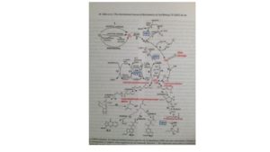
Fig.1. – Valko, M., et al. (2007). “Free radicals and antioxidants in normal physiological functions and human disease.”
https://www.sciencedirect.com/science/article/abs/pii/S1357272506002196

Some important questions for your to consider:

  • Do you think your healthcare provider understands the chart above? I barely do and I have been studying this intensely for 3+ yrs!
  • Can you see how high-dose iron supplements, or worse yet, iron infusions might bring those two diagrams to life, especially the second one?
  • Can you see how measuring a single, controversial and metabolically inactive protein, ferritin, is not a proper, relevant, appropriate or complete measure to assess the complexity of iron metabolism?

What I would offer up as a key companion article to Valko, 2007 is an important study that I have shared before by Sir Douglas B. Kell, PhD entitled “Iron Behaving Badly”

Kell, D.B. (2009). “Iron behaving badly: inappropriate iron chelation as a major contributor to the aetiology of vascular and other progressive inflammatory and degenerative diseases.”
https://pubmed.ncbi.nlm.nih.gov/19133145/
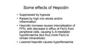
Research Gate image
https://www.researchgate.net/figure/Some-effects-of-hepcidin-summarizing-the-fact-that-hypoxic-condition-can-suppress-it-and_fig8_23767901

I wonder how many have read the Douglas Kell article above? The difference between these two compelling studies is that Valko, et al., keep the illusion alive that oxidative stress can come from many metabolic sources. 

Whereas Professor Kell puts the spotlight squarely where it belongs which is rogue iron found in the ferritin molecule. There was no pretense in his accomplishment of research he has done. 

Know that there is 60x more iron than copper in the human body is very telling as stated by Gutteridge and Halliwell in their 1994 book.

Gutteridge, J.M.C., Halliwell, B. (1994). “Antioxidants in nutrition, health and disease.”


Then a bright member of the Magnesium Advocacy Group, Jim Urban, shared this expressive and penetrating piece on the toxicity of iron which I encourage you all to read:

Zacharski, L.R. (2014). “Ferrotxic Disease: The Next Great Public Health Challenge.”
https://academic.oup.com/clinchem/article/60/11/1362/5621712

If you really want to explore these iron issues and dynamics, here is the Full Monty Iron blood test that I routinely recommend to accompany the HTMA:
https://therootcauseprotocol.com/order-lab-tests/

The key is then getting it properly interpreted by someone who knows the difference between:

  • Anemia of Chronic Inflammation which is quite common and caused by a state of excess iron that is in hiding in tissues, organs etc. 
  • Anemia of Iron Deficiency, which is next to impossible on a planet that has 36% of its composition, made up of iron.

I would like you to fully understand what Anemia of Functional Iron is really all about.  Iron is supposed to be recycling not being stored. The understanding of iron status is so important especially if it is based on one marker, ferritin that is not telling the full story.

A votre sante!

MORLEY M. ROBBINS

For Facebook Discussion:
www.facebook.com/groups/MagnesiumAdvocacy/permalink/1126634560737945

Iron Toxicity Post #41: Anemia of Inflammation and how it relates to anemia

Iron Toxicity Post #41: Anemia of Inflammation and how it relates to anemia

In my ever-continuing efforts to get folks from the entire world over to restore and protect their magnesium (Mg) status, I’ve taken a most unexpected and extended journey into and through the wacky, Byzantine of the most revealing world of iron research. Fortunately, this detour has enabled us to better understand why we are all so compromised on our Mg status, and consequently on our health.

Now mind you, today’s post is not what I had drafted yesterday. I had assembled a set of “killer” research studies, had excerpts highlighted, had the entire post written and then lost the internet connection at the hotel where I was receiving some training in essential oils. Are you kidding me! (I took the set back in stride as I figured that there was something “missing” in my message)

And indeed there was!

I’ve decided to honor Einstein, once again, and seek to simplify this “You ain’t iron anemic!” message, once more.

Think of this as remedial ironology 101.

First some of the key fundamentals:

  • Definition of anaemia, which is from Greek: “an” meaning without and “haima” meaning blood.  This was adopted in the early 19th century from modern latin as a condition marked by a deficiency of red blood cells or of haemoglobin in the blood, resulting in pallor and weariness.
  • Unbound (free) iron has the unique ability to bring about 3 critical and damaging developments within the cells:
    1. Iron lowers pH and forces the cell to an acidic pH.
    2. Despite hemoglobin’s known role to deliver oxygen, unbound free iron lowers oxygen in the cell (low pH = low oxygen; it’s a basic chemical and physiological property on this planet, but know that optimal oxygen is at pH = 7.4, not at the other end!)
    3. Iron uncouples oxidative phosphorylation and lowers the production of ATP inside the mitochondria (one study that I read indicated that it was as much as a 96% loss of ATP!) 
  • Ferritin is not an indication of haemoglobin status and therefore is not even close to being an accurate assessment of oxygen-transport status, which is actually hemoglobin’s job.  Ferritin is an iron storage protein, not active indicators of iron neither embolism nor iron physiology. A ferritin only test is a faulty and misleading indication of iron status! (Douglas Kell PhD’s article “Iron behaving badly” in 2009 has put the exclamation point on that issue.)
  • It has been a well-known fact, since the 1860’s that iron anemia is a clinical indication of copper deficiency.  We refer copper deficiency as low bioavailable copper which has a role in the creation of:
  • Erythropoietin (EPO) in the adrenals (a hormone to signal for new red blood cells in the bone marrow)
  • The production of heme 
  • The insertion of iron into heme
  • The creation of haemoglobin (a ring of 4 heme proteins)
  • The proper use and impact of heme oxygenase 1 to recycle iron

Again, all of the above depends on bioavailable copper.

  • Low ferritin is a definitive sign of low bioavailable iron, thus iron dysregulation and not iron deficiency.
  • Ferritin comes in two types of chains, ferritin-heavy, which is very dependent on ceruloplasmin activity and ferritin-light, which lacks ceruloplasmin activity.

This brings about the question why does practitioners all over the world administer iron, given that it will only cause the oxygen levels to drop even further.  The fact that excess, unbound iron lowers pH and not one single word is ever mentioned about copper status or ceruloplasmin status or the status of B-vitamins that are very much a part of the iron metabolism.

I invite you to read a wonderfully written & researched article by Ray Peat, PhD, entitled “Iron’s Dangers”:
https://therootcauseprotocol.com/wp-content/uploads/2023/02/Irons-Dangers-Ray-Peat.pdf

Those that have followed me for any length of time know that I have an intense love/hate relationship with this gifted, enigmatic nutritionist. This would be one of my favorite posts that he’s written and possibly because he, too, is sounding this iron alarm. One of my favorite lines from this article:

”Iron deficiency anemia does exist, in laboratory situations and in some cases of chronic bleeding, but I believe it should be the last-suspected cause of anemia, instead of the first.”

I also will highlight his summary:

Iron is a potentially toxic heavy metal; excess can cause cancer, heart disease, and other illnesses. 

Other heavy metals, including lead and aluminium, are toxic; pans and dishes should be chosen carefully. 

Iron causes cell aging. [This is important]

Drinking coffee with iron rich foods can reduce iron’s toxic effects. 

Use shrimp and oysters, etc., to prevent the copper deficiency, which leads to excess storage of iron. 

Avoid food supplements, which contain iron.

Take about 100 units of vitamin E daily; your vitamin E requirement increases with your iron consumption.”

I am not buying the you are anemic façade so what do I think all of these very low ferritin’s, low serum iron’s and low % saturation’s is really all about?

All signs and research are pointing toward Anemia of Inflammation (AI), which I will delve into with rigor and compelling studies to put this ferritin-driven-anemia to rest.

All is not as it seems, but it’s getting clearer and clearer.

A votre sante!

MORLEY M. ROBBINS

For Facebook Discussion:
www.facebook.com/groups/MagnesiumAdvocacy/permalink/1081365178598217/

MAG-pie Alert!… #37 TOXICITY OF IRON

MAG-pie Alert!… #37 TOXICITY OF IRON

MAG-pie Alert!… #37 TOXICITY OF IRON13566995_10154916251022908_7591494815194509420_n

MAG-pie & MAG-net Alert
For those who wonder WHY I wear a “Tin Hat” ALL DAY LONG…
We’re coming up on 25 years of suppression & deception, folks…

A votre sante!
http://www.stage2omega.com/united-states-should-be-on-this-list-why-the-heck-not/
Iron Toxicity Post #40: Medication can cause iron induced oxidative stress!

Iron Toxicity Post #40: Medication can cause iron induced oxidative stress!

This is an article about how Pharmaceuticals can affect the mitochondria causing collateral damage, which then creates disorders such as schizophrenia, diabetes, PD, CFS, and non-alcoholic steatohepatitis.
www.sott.net/article/321987-Thanks-Big-Pharma-for-the-Mitochondrial-collateral-damage

Anytime you see the phrase reactive oxygen species (ROS) or its equivalent, start seeing the destructive nature of iron-induced oxidative stress, which also implicate disorders such as schizophrenia, diabetes, PD, CFS, and non-alcoholic steatohepatitis. 

Please review the 72 most popular drugs and their impact on the energy centers (mitochondria) in our cells.

A votre sante!

MORLEY M. ROBBINS

For Facebook Discussion:
https://www.facebook.com/groups/MagnesiumAdvocacy/permalink/1068813699853365/

Iron Toxicity Post #39: excess unbound iron is very bad! Get properly tested.

Iron Toxicity Post #39: excess unbound iron is very bad! Get properly tested.

(Formerly ITP#39)

Please share this “oldie, but goodie” with your favorite practitioner:

Williams, D.M., Lee, G.R., Cartwright, G.E. (1976). “Role of Copper in Mitochondrial Iron Metabolism.”
https://ashpublications.org/blood/article/48/1/77/160532/Role-of-copper-in-mitochondrial-iron-metabolism

I am also quoting from another article that I was just reading: 

Griffith, D.P., et al. (2009). “Acquired Copper Deficiency: A Potentially Serious & Preventable Complication Following Gastric Bypass Surgery”
https://www.ncbi.nlm.nih.gov/pmc/articles/PMC2712481/

…Mitochondrial cytochrome c oxidase, which plays a critical role in the transfer of Iron to the cytosol for incorporation into heme [23]. Ceruloplasmin ferroxidase is also thought to contributes to anemia, as it is essential [emphasis added] in the loading of transferrin with iron in the liver and is markedly diminished in parallel with copper (ceruloplasmin) [24].” (Please note, the former linked article is citation [23] noted above)

It is well known in iron research circles that low hemoglobin is a clinical sign of copper deficiency (i.e. low bioavailable copper). I will now go out on a limb and assert that low ferritin, low serum iron, and low % saturation are indicative of copper deficiency, as well. 

What is clearly emerging from the literature is that these 3 markers noted are highly correlated and highly indicative of what is called Anemia of Chronic Illness, or Anemia of Inflammation.

They are not indications of iron deficiency. They are signs of iron dysregulation that is fueled by low bioavailable copper as best expressed by low ceruloplasmin.

For those that want to see it in “red,” please order this test:
requestatest.com/mag-zinc-copper-panel-with-iron-panel

What I am finding is that you cannot properly interpret a set of iron markers without knowing magnesium RBC, plasma zinc, serum copper and serum ceruloplasmin. 

It is essential to have an understanding of the minerals that regulate iron, in addition to iron itself.

A votre sante!

MORLEY M. ROBBINS

For Facebook Discussion:
https://www.facebook.com/groups/MagnesiumAdvocacy/permalink/1065612353506833/