Iron Toxicity Post #47: The truth about the cause of mitochondrial dysfunction (as least as I see it)

Iron Toxicity Post #47: The truth about the cause of mitochondrial dysfunction (as least as I see it)

This post is about the truth of the cause of Mitochondrial Dysfunction, at least as I see it.

Over the years, there have been certain phrases that have chaffed me, among the most offensive is this term: “Mitochondrial Dysfunction.” 

It is a term that conjures up an equal amount of fear, wonderment and confusion like that often used phrase; “Oh, you have a lesion” 

There are thousands and thousands of articles, both for the layperson and the scientist, written about this mitochondrial condition that is rapidly becoming a pandemic on this planet. 

When all the dust settles, it’s not unlike writing a Magnus Opus about how to drive a car, with meticulous detail to every facet of that automotive experience with the notable exclusion of two key and fundamental dimensions to a successful drive:

  • Forgetting to tell you that it’s really important to bring a key to start the engine
  • Forgetting to tell you that the engine must have fuel to keep it running

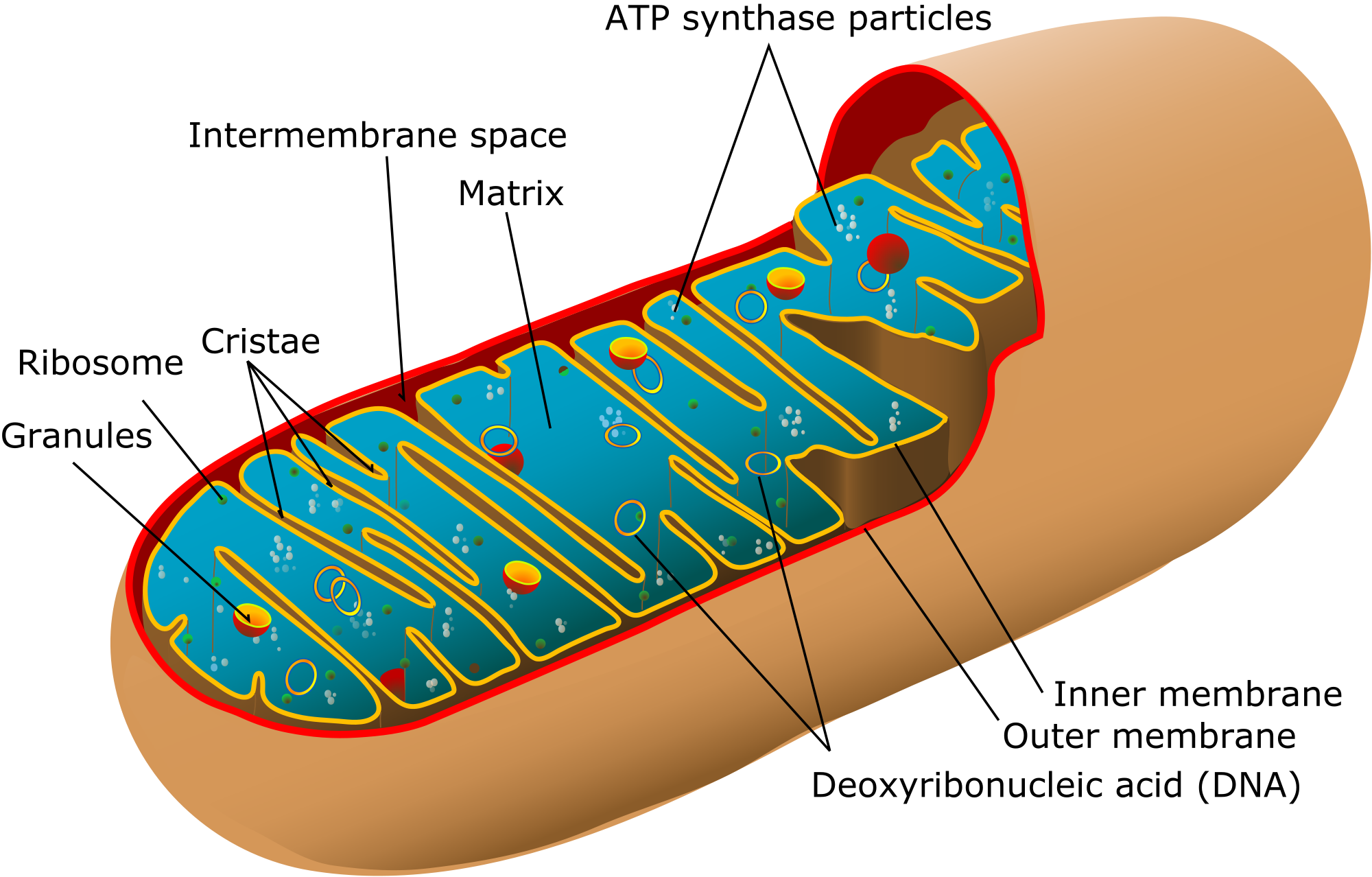
Please take a good long look at the picture below.

Figueiredo, P.A, et al. (2008). “The role of mitochondria in aging of skeletal muscle.”

https://www.researchgate.net/figure/ROS-release-by-the-electron-transport-chain-ROS-are-released-into-the-mitochondrial_fig1_5675258

I’ve been looking for this type of picture that dumbs down the mitochondrial electron transport chain (ETC) so that I can actually understand it!  What I love is that this diagram helps to fill in many blanks that are ubiquitous and cleverly disguised in so much of the scientific literature.

The mitochondria are where our adenosine tri-phosphate (ATP) gets made.  ATP is the packet of energy that fuel our cellular, tissue, organ and bodily functions. 

This takes place at Complex IV of the ETC. It’s ridiculously important. The mitochondrial cytochrome proteins that make up these complexes are the most extensively studied electron-transfer proteins.

The step that should make you sit up and take special notice is called cytochrome c oxidase (Complex IV) because it is here that four electrons get transferred to oxygen so that water (H2O) can be made so the ATP molecule can be re-made from ADP. As it turns out these cytochromes are actually heme proteins, which means that they contain ferrous (Fe2+) iron, which is the most reactive form of iron. This critical cytochrome c oxidase enzyme does not work without bioavailable copper, which is the key to make the ATP- engine work.

Who do you think is supplying the copper energy for this critical aspect of our metabolism? Ceruloplasmin! For those seeking mineral insight into this metabolic dynamic, please read Broderius, 2010:

Broderius, M., et al. (2010). “Levels of plasma ceruloplasmin protein are markedly lower following dietary copper deficiency in rodents.”

 https://www.ncbi.nlm.nih.gov/pmc/articles/PMC2854028/pdf/nihms-192848.pdf

Also note that there’s oxidative stress being given off on both sides of this mitochondrial process. Oxidative stress is a natural byproduct of these activities.  It turns out the greatest source of endogenous oxidative stress occurs across these critical mitochondrial actions of Complex I through to V.

It is common to think that only copper and zinc superoxide dismutase (Cu/Zn-SOD) or known as SOD I, works the intracellular space (i.e. outside the mitochondria) and that only manganese superoxide dismutase (Mn-SOD) or known as SOD II, works the intra-mitochondrial membrane (i.e. inside the mitochondria). I used to think that was the case, too, until I found this ancient blockbuster (pdf.) article on ceruloplasmin (Goldstein, 1979):

Goldstein. I.M., et al. (1979). “Ceruloplasmin.”
https://www.jbc.org/article/S0021-9258(18)50692-X/pdf?fbclid=IwAR0S9X_gQrsWmZ-E4zW4cEfbYl6plv-LvT0IqlAx1AaSYYY0SA3airR3StI

What’s notable about the ceruloplasmin ferroxidase enzyme is that it is a recognized scavenger of superoxide radicals. They are those reactive oxygen (*O2-) molecules that are quite destructive. As you can see from the diagram above, they can and do devolve both inside and outside the mitochondria. They turn into the most destructive molecule in the body hydroxyl radical (OH-). This is the origin of what dings deoxyribonucleic acid (DNA) to create epigenetic chaos to support methylenetetrahydrofolate reductase (MTHFR).

This destructive molecule also leads to a breakdown in the efficiency and impact of the energy production of these mitochondria, which is known as chronic fatigue syndrome (CFS) as well a host of other chronic conditions of low energy.

You can now see how this destructive molecule also disrupts the cellular machinery of our body systems such as livers, hearts, brains, kidneys, joints, and muscles.

Now, can you all begin to see why this thing mitochondrial dysfunction is such a big deal, some cells have as many 200-500 mitochondria in them but I believe it is not nearly as complicated as it is methodically made out to be. 

Furthermore, what I was not certain about until today is that ceruloplasmin is not just restricted to the cytoplasm (i.e. outside the mitochondria). It turns out that this scavenger is, for a fact, found inside the mitochondria, as well. It is well documented in these Russian researches!

Vasin, A.V. et al. (2005). “Mitochondrial ceruloplasmin of mammals.”
https://www.ncbi.nlm.nih.gov/m/pubmed/15773547/

Balevska, P., et al. (1975). “Studies on the transfer of copper from ceruloplasmin to mitochondria.”
https://www.ncbi.nlm.nih.gov/m/pubmed/1232838/

I have learned over the years to regard the abstracts that have no abstracts as being really important.

The plot begins to thicken with the benefit of these penetrating insights and here’s what I think is the blockbuster.

It turns out that 20% of the mitochondrial bi-lipid membrane is made up of a phospholipid called cardiolipin. While it is 20% of the total composition, it is actually 50% of the phospholipids found to be a part of the ETC (i.e. Complexes I – V) lipids. Here is the article that offers keen insight to the dynamics between cardiolipin and superoxide:

Soussi, B., et al. (1990). “H-n.m.r. evaluation of the ferricytochrome c-cardiolipin interaction: Effect of superoxide radiacals.”

https://www.ncbi.nlm.nih.gov/pmc/articles/PMC1136634/pdf/biochemj00192-0227.pdf

It turns out that cardiolipin is essential for the function of cytochrome c oxidase. Wow! I have read scores of articles on this Complex IV enzyme, cytochrome c oxidase, but I have never read that before. Their interaction is electrostatic in nature, as I’m sure everything is that happens in our bodies.

So what does this mean?

  • Copper is the key to start this cytochrome c oxidase enzyme production of ATP
  • Cardiolipin is the fuel upon which the cytochrome c oxidase enzyme works

What do the professionals from Dover have to say in the summary about the origin of mitochondrial dysfunction:

“Mitochondrial dysfunction… is related to a decreased cytochrome c oxidase activity, ultimately because of cardiolipin peroxidation [i.e. rusting of the fats] (Smith et al, 1980; Okayasu et al, 1985) causing a diminished cytochrome c binding to this enzyme.”

So in the absence or the presence of lowered production of ceruloplasmin, those ubiquitous superoxide (*O2-) molecules get further radicalized by ferrous (Fe2+) iron. Then into the OH- radical that then oxidizes the cardiolipin.

I happen to think it is relevant and most vital that ceruloplasmin brings both the copper (key) and ensures that the cardiolipin (fuel) does not get rusty as we talked about the car analogy earlier in the post.

Ceruloplasmin, as the Ferroxidase enzyme, converts ferrous (Fe2+) iron into ferric (3+) iron, the latter form of iron being what can then be used in cellular transport and other metabolic proteins to keep us healthy and energized. Isn’t it amazing that ceruloplasmin does this and more?

Even though this term mitochondrial dysfunction seems hopeless and it is a critical issue we all recognize and know. It can be whittled down to size and made far easier to comprehend and understand the why and how, and then we can prevent or reverse its development in our metabolism.

It is not a Medical Disease. Mitochondrial dysfunction is simply a clinical sign of Mineral Dysregulation – pure and simple.

Those who are seeking direction on what to do to address this, you can go here and get started: https://therootcauseprotocol.com

A votre sante!
MORLEY M. ROBBINS

For Facebook Discussion:
www.facebook.com/groups/MagnesiumAdvocacy/permalink/1209061335828600/

Iron Toxicity Post #46: The “Reader’s Digest” version of what causes mitochondrial dysfunction

Iron Toxicity Post #46: The “Reader’s Digest” version of what causes mitochondrial dysfunction

This is the “Readers Digest” version of what causes mitochondrial dysfunction.

I am following this up more information about this topic in the next iron toxicity post.

=> Metabolic Derangement
is caused by

=> Mitochondrial Dysfunction
which is caused by

=> Mineral Dysregulation
which is caused by 

=> Our Mineral Deficient Diet, which cause diagnoses and drugs as a result which are caused by, respectively

Intriguing, eh?

A client/colleague recently shared with me that Hippocrates was forever seeking

… The cause
… of the cause
… of the cause…
Well, now we know the origin of all of our symptoms.

Please know, the path back to optimal health and metabolic homeostasis is outlined here: https://therootcauseprotocol.com/

A votre sante!
MORLEY M. ROBBINS

For Facebook Discussion:
https://www.facebook.com/groups/MagnesiumAdvocacy/permalink/1209045499163517/

Iron Toxicity Post #45: Brain aging and Alzheimer’s

Iron Toxicity Post #45: Brain aging and Alzheimer’s

This post is about brain aging and Alzheimer’s.

Attached to this post is from a penetrating study on the metabolic origin of brain aging and Alzheimer’s.

Atamna, H., et al. (2002). “Heme deficiency may be a factor in the mitochondrial and neuronal decay of aging”
https://www.ncbi.nlm.nih.gov/pmc/articles/PMC137500/pdf/pq2302014807.pdf?fbclid=IwAR30u0XbRCdgGmUT8lbwGbko1v1f44K8WqXlrxp7yhuEnZXGNmXlWXjrhwI

Trust me when I say this, Alzheimer’s disease (AD) is no disease!

Given that this neurological condition is ravaging society and our bank accounts around the globe, I would strongly encourage you to study this carefully.

The bottom line, this condition stems from Heme deficiency that is the result of dysfunction in one enzyme called ferrochelatase. It contains what are called iron-sulfur clusters, but what is missing in this otherwise scintillating study is the fact that ferrochelatase is dependent on bioavailable copper, aka ceruloplasmin.


Furthermore, note that iron deficiency does not mean a lack of iron, but actually means a shortage of functional iron!  And we all now know that it’s copper that enables iron to work properly hence known as copper<>iron metabolism. This means it is a ventriloquist act and iron is the dummy.

What is especially important to understand is that when heme does not get made properly, there are a series of downstream metabolic impacts, not the least of which is iron accumulation in the brain. Lack of functional iron creates a metabolic requirement to store Iron. 

Share this research with your loved ones, friends and their healthcare providers who may not be aware of this critical mineral dimension. This is due to misleading and lacking clinical training. 

A votre sante!
MORLEY M. ROBBINS

For Facebook discussion:
https://www.facebook.com/groups/MagnesiumAdvocacy/permalink/1182486528486081/

Iron Toxicity Post #44: Iron Elephant

Iron Toxicity Post #44: Iron Elephant

We learned this from Jill Cohen-Wilson that the “lamp of magnesium” is starting to gain traction — in her doctor’s office in Long Island, NY. Wow, even I’m impressed!

And in fitting tradition, Dr. Liz & I discovered that today’s quotation from Rumi was all about this dynamic of being in the dark.
https://www.goodreads.com/quotes/117359-some-hindus-have-an-elephant-to-show-no-one-here

I am reminded of the famous story of a group of people, who are blindfolded (i.e. in the dark), being asked to describe an elephant from each of their unique perspectives, as is cleverly depicted in the cartoon below.

So what is my point? 

We have all been blind to the iron elephant in our body without the “light” of the research and insights that we are learning and sharing here at Magnesium Advocacy group (MAG): 

These symptoms, conditions, syndromes and dare I say, these diseases appear very real and very sinister. Why?  We have been blind to the ravaging impact of iron toxicity and its generation of oxidative stress in our bodies. Well, fortunately, those days are drawing to a close as we share this truth more widely.

For those seeking to learn more about these iron dynamics and their metabolic impact in our bodies, please read this wonderful book by Roberta Crawford who runs a NFP dedicated to educating the public about iron, and iron loading disorders, entitled “The Iron Elephant”. It’s a wonderful book and well worth your time to read.

Crawford, R. (2001). “The Iron Elephant”
https://www.amazon.com/Iron-Elephant-Should-Danger-Excess/dp/0963254774

Again, our sponsor magnesium is, indeed, the “Lamp of Life”

A votre sante!
MORLEY M. ROBBINS

For Facebook Discussion:
https://www.facebook.com/groups/MagnesiumAdvocacy/permalink/1172056309529103/

Iron Toxicity Post #43: The ‘gestation and birth’ of iron toxicity

Iron Toxicity Post #43: The ‘gestation and birth’ of iron toxicity

Today is my Natal Day! Let me first thank the many folks who’ve taken the time to reach out and celebrate with me. I’m most grateful to this loving and giving community, and I’m also grateful to my parents, especially my Mom, for allowing this little life to exist.

My Mom’s Dad, John M. deLashmutt, Jr. had an annual tradition of giving a gift to his family on his natal day. That always left a big impression on me. So given that Magnesium Advocacy Group (MAG) is a second family of sorts for me, well here’s my gift to you all. 

It’s a bit complicated, a bit convoluted, and also a bit counter-conventional.

I realize that I have been an incessant bore this year with my non-stop posts about iron toxicity.  Well, we are not done yet. Today’s post is intended to drill into how this iron chaos starts, from the very inception of life. 

I am piggybacking on a recent post by a member who shared some illuminating research recently (Steer, et al, 1995), but only four people noted its existence last week. 

I believe this contemporary issue of “anemia of pregnancy” is completely out of control. It is completely misleading by clinical training as well as understanding of basic mineral metabolism especially as it relates to haemoglobin (Hgb) & red blood cells (RBC). 

It turns out that among the most important aspects of early life, as a mammal, is creating and managing our red blood cells.

Fraser, S.T., et al. (2011). “Heme Oxygenase-1: A Critical Link between Iron Metabolism, Erythropoiesis, and Development.”
https://www.hindawi.com/journals/ah/2011/473709/?fbclid=IwAR1Byk4YeUFf7eutIUxwEtGnCshaYVhW1ylMENrPGATsIKolwMxiRpem0WA

An important quotation from the very beginning of this article was:

“The first mature cells to arise in the developing mammalian embryo belong to the erythroid [Red Blood Cell] lineage. This highlights the immediacy of the need for red blood cells during embryogenesis and for survival.”

For those that have been following my incessant posts these past twelve months, you will know that every facet of red blood cell formation and metabolism is copper dependent. It not about iron status, as we know.

Fox, P.L. (2003). “The copper-iron chronicles: The story of an intimate relationship.” (No full text available now)
https://www.ncbi.nlm.nih.gov/pubmed/12572662

I would encourage you to read this classic article as it explains how and why this has become so confusing.  What the Fraser (2011) article also highlights is yet another aspect of Hgb metabolism is copper dependent, which is heme oxygenase-1 (HO-1) protein that is the rate-limiting enzyme in the breakdown of haemoglobin.

Ideally, these RBCs that carry the Hgb that carry the oxygen are supposed to live, on average around 120 days and then they get recycled. As it turns out, that is when in copper deficiency state, the amount of HO-1 increases thus causing an increase in the rate of Hgb turnover.

Johnson, W.T., et al. (2004). “Increased heme oxygenase-1 expression during copper deficiency in rats results from increased mitochondrial generation of hydrogen peroxide.”
https://www.ncbi.nlm.nih.gov/m/pubmed/15173392/

This is not an insignificant fact about RBC/Hgb metabolism.

I would go so far as to state that the research that the member brought to our attention especially the Steer, et al., 1995 in BMJ, is among the most powerful studies to explain how and why society is being methodically exposed to excess iron.

Steer, P., et al. (1995). “Relation between maternal haemoglobin concentration and birth weight in different ethnic groups.”
https://www.ncbi.nlm.nih.gov/pmc/articles/PMC2548871/

The magnitude of this article is that Hgb in a pregnant woman, especially in the second half of the pregnancy is supposed to drop to 8.5-9.5 mg/L. 

This is a critically important fact and set of studies that challenges the conventional clinical paradigm of where iron levels are supposed to be during the course of a healthy pregnancy.

What they also found is that as Hgb drops, birth weight of the infant rises. High Hgb is a sign of potential premature birth and pregnancy risk.

What is the big deal about adding iron to a woman who’s Hgb appears low?

  • It’s a clear signal that more likely than not that the pregnant woman and the babino are copper deficient.
  • It increases the chances of undermining the production and metabolism of ceruloplasmin that is key for proper healthy copper metabolism.
  • It increases the chances for oxidative stress in both the Mom and developing foetus.  A particularly poignant and penetrating look at this dynamic was outlined by Keyer & Imlay (1997):

Keyer, K., Imlay, J.A. (1997). “Inactivation of Dehydratase [4Fe-4S] Clusters and Disruption of Iron Homeostasis upon Cell Exposure to Peroxynitrite*”
http://www.jbc.org/content/272/44/27652.full.pdf

This research should send a chill down the spine of those taking iron supplements and those getting iron infusions. 

I encourage any who are pregnant, or planning to get pregnant to read and study this research carefully. Please share it liberally with your Obstetrician, Midwife, etc. and do not be surprised that they may not know these issues, understand these implications, nor practice in a way to avoid iron impacts.

I would encourage you to have the Full Monty Iron panel and have it properly interpreted.
https://therootcauseprotocol.com/order-lab-tests/

A votre sante!
MORLEY M. ROBBINS

For Facebook Discussion:
www.facebook.com/groups/MagnesiumAdvocacy/permalink/1169503143117753/