Iron Toxicity Post #52

Iron Toxicity Post #52

In my continuing efforts to “take off the gloves,” about how they are being poisoned by iron fortification, iron supplementation, iron infusions, iron stimulation, iron mesmerisation, etc. Today’s post will present some new dimensions to this iron discussion that will hopefully “stimulate your little grey cells”, as the great fictional and enigmatic detective, Hercule Poirot, so frequently said while solving equally perplexing, yet elusive, murder mysteries.

One of my clients suggested that I might find this blog on curcumin and iron useful:
margaret.healthblogs.org/life-with-myeloma/discovery-of-curcumin/curcumin-and-iron/

In fact, it’s quite good, it’s well written, but it is quite misleading.  Nevertheless, this blog rocked my world, and I am grateful to Margaret for that!

I’ve read a couple hundred articles on iron, iron metabolism and iron dysregulation. I also read my fair share of articles on ferritin, and yes, I knew that ferritin came in two forms: an active state (heavy-ferritin with ferroxidase function) and an inactive state (light-ferritin without this enzyme function). I hadn’t shared that bombshell as of yet, but did any of you know that ferritin had two forms?

I really wonder now, are we low in the active or the inactive state of ferritin?

But what this Blogger, Margaret, wrote that really caught my eye is the fact that ferritin has a relationship to both iron and oxygen!

Oxygen? How, exactly, did I miss that functional factoid? Given that ferroxidase has a special relationship with oxygen, wouldn’t it be best to have the heavy (active) ferritin form? I wonder how many healthcare providers are aware of these important iron dynamics?

Liu, X., et al. (2006). “Iron at the center of ferritin, metal/oxygen homeostasis and novel dietary strategies.”
www.scielo.cl/pdf/bres/v39n1/art18.pdf

Theil, E.C. (2003). “Ferritin: At the Crossroads of Iron and Oxygen Metabolism.

It turns out that iron is actually a Janus – it wears two faces across numerous dimensions. Here are some critical and dualistic facts about iron:

  • Iron comes in two oxidative states: ferrous (Fe2+, soluble) vs. ferric (Fe3+, insoluble)
  • Ceruloplasmin comes in two forms: ferroxidase enzyme vs. immuno-reactive protein
  • Ferritin, as noted above comes in two forms: H-ferritin (active) vs. L-ferritin (inactive)
  • Iron has two roles with oxygen: carrier of oxygen (Hemoglobin) vs. creator of oxygen radicals (reactive oxygen species)
  • Iron dysregulation comes in two forms: “anemia of iron deficiency” (very rare!) vs. “anemia of chronic inflammation” which I believe is pandemic on this planet
  • Iron metabolism has two dimensions: Iron metabolism vs. Copper<>Iron metabolism
  • Copper has two oxidative states, as well: cupric (Cu+) vs. cuprous (Cu++)

Now you can understand how I feel about iron anemia vs. iron dysregulation and practitioners’ use of high and low to describe our iron status is very misleading!

The worlds of medicine, nutrition and mineral metabolism are littered with this “High” vs. “Low” mentality. It is criminal, and it is the very origin of how we’ve gotten so confused about iron and have become so iron toxic!

There is no such thing as iron metabolism.

For a fact, it is copper<>iron metabolism, the leading researchers know this, and point out that these two metals are joined at the hip of ceruloplasmin (Cp). This Cu<>Fe metabolism is actually a Ventriloquist Act, which means iron is the dummy. 

It is not all about how much iron; it’s all about how usable is it. We are learning that ferritin can be most unusable especially when ferritin is in the Light-ferritin form.

I have come to reflect on it, ferritin is the ultimate Trojan horse, especially in a body that lacks ferroxidase function in its ceruloplasmin, as well as in its ferritin.

We all know that horses are most useful when they are functional? Well, this is true of iron, and ferritin, too!

The inactive “Trojan ferritin” being fed more iron is simply adding insult to your non-functional iron pool, especially if your mag RBC and serum ceruloplasmin are both anemic. 

So, if you still have a mind to think that you’re “anemic”, even after all this post. Please take this little test to assess your indications of iron knowledge. 

  • Do you still think anemic only means a lack of iron?
  • Do you still believe your healthcare provider understands the Janus duality of iron, as noted and outlined above?
  • Do you still use the redundant terms “high” and “low” to describe your dysfunctional iron and your dysfunctional ferritin status?
  • Do you still not understand what “anemia of chronic inflammation” is referring to?
  • Have you still not gotten the Full Monty Iron panel, nor had it properly interpreted?
  • Have you still not taken the time to view, nor reflect on the Root Cause Video?
  • Have you still not started to adopt the Stops and Starts of the Root Cause Protocol?
  • Have you still not stopped taking multi-vitamins/iron supplements/iron infusions?
  • Do you still continue to research what the signs and symptoms of “anemia” are on the Internet?
  • Have you still not read any of the 50+ posts on iron toxicity?
  • Have you still not questioned your doctor’s understanding of the Janus duality of iron?

If you say yes to any of the above questions, then you are still following iron anemia rather than iron/copper metabolism.

What I am noting in more and more iron research articles, are increasing references to the term “Iron deficiency” being openly referred to as the “lack of functional iron”. Not an absolute “deficiency of iron!” Big difference! 

Copper<>Iron metabolism is all about making the iron functional. That is only possible with the presence and prevalence of copper and copper-driven enzymes like ceruloplasmin (Cp). Also the copper dependent anti-oxidant enzymes (SOD, CAT, & GPx) designed to neutralize the iron-driven reactive oxygen species (ROS) that unbound iron creates with no limits within our bodies, our tissues, and our cells.

If your healthcare practitioner(s) are not questioning your true iron status and are not focusing on your magnesium and ceruloplasmin status. Then treating you with calcium, hormone-D, zinc and iron. And worst of all, are still “attacking” your alleged “copper toxicity,” then they are not addressing your body’s core needs or honoring its innate healing potential.

This duality of iron is very poorly understood! The systemic simplistic solution of adding more iron to your iron dysfunction is both one-sided and dangerous.

Here is a great way to start the process of better understanding your iron status, as well as supporting its recovery and rebalance within your body:
https://therootcauseprotocol.com/about/
A votre sante!
Morley M Robbins

For Facebook Discussion:
https://www.facebook.com/groups/MagnesiumAdvocacy/permalink/1261433993924667/
Iron Toxicity Post #51: The Deceit of ‘Anemia’

Iron Toxicity Post #51: The Deceit of ‘Anemia’

This is dedicated to members and other clients, as well as the millions of women/moms around the globe who are being misdiagnosed due to the misinformation of iron metabolism.

This post, the gloves are coming off while I addressed the common iron anemia thing. 

If any of the following conditions apply, I would sincerely ask that you read this post and the accompanying articles, very slowly and carefully:

  • “I’m anemic…”
  • “My iron is low…”
  • “My ferritin is too low…”
  • “I’m tired and I need more iron…”
  • “I need to raise my ferritin more…”
  • “I’m taking iron supplements/infusions…”
  • Or any variation on these Iron themes.

You are not “anemic!

The ability or the know how to measure iron properly in the human body takes into account that every facet of iron activity is ruled and regulated by bioavailable copper. 

If you are at all curious about my stance and wonder why I believe that  the current understanding about iron metabolism is misguided, please read this important article below:

Messner, D.J., Kowdley, K.V. (2010). “Biting the Iron Bullet t: Endoplasmic Reticulum Stress Adds the Pain of Hepcidin to Chronic Liver Disease.” Hepatology; 51(2):705-707
www.ncbi.nlm.nih.gov/pmc/articles/PMC2849800/pdf/nihms180510.pdf

What this article is trying to tell us is:

“Inflammation and iron are known [emphasis added] extracellular stimuli for hepcidin expression.”

What does that mean?

Well, hepcidin (Hpn), which has 25 amino acids, is considered in today’s conventional view to be  the regulatory hormone for iron metabolism. But when you compare Hpn to ceruloplasmin, which has 1,046 amino acids, you can begin to understand how it dissociates in the teaching. 

This article really highlights what is really going on with getting iron out of the cell and back into circulation: 

Musci, G., et al. (2014). “Ceruloplamsin-ferroportin system of iron traffic in vertebrates.”
www.ncbi.nlm.nih.gov/pmc/articles/PMC4050113/
The failure to understand the pivotal dynamics between Ceruloplasmin <> Ferroportin <> Hepcidin leaves us with practices that don’t address the body’s core needs or honor its innate healing potential.

Iron metabolism is meant to be a sophisticated recycling program.  Iron is meant to be in constant circulation, literally, and ceruloplasmin & ferroportin guarantees that process. What happens is increased expression of hepcidin is disrupting this process therefore more hepcidin is not what you want rather you want more ceruloplasmin to get the iron moving.

So, when iron is showing low on the blood work, it is not unreasonable to assume that a pathogenic infection has triggered our immune system to do what it has been designed to do. It pulls all of the iron out of the blood and put it into storage. Why does it do that? 

All critters (bacteria, virus, fungus and parasites) must have iron to live, to replicate and to thrive. Our immune system is finely tuned since life to keep that iron from them. This system has worked for a long time, until changing our food and giving Rx meds began around 75yrs ago. What happened is that it has changed the foundational understanding of copper, as well as the critical interplay between Copper <> Iron in regulating these metals. This has an impact on our metabolic functions, especially in our liver.

Ok, I get that, so then what’s the problem?

Well, as laid out in this Messner (2010) article, we now are learning that inflammation and iron increases the production and expression of hepcidin!  Remember that we know that iron causes inflammation.

So what does that mean?

Increased Hepcidin => 

Increased Iron Storage => 

Decreased iron appearance in blood work

To translate that key phrase, noted earlier, from this Messner (2010) article to:

“Inflammation and iron are known extracellular stimuli for increased iron storage [emphasis added] which causes iron to show up low on ferritin, show up low on serum iron, and show up low on % saturation markers.”

 Unfortunately, Hepcidin cannot be ordered from commercial labs, so it rarely gets measured.  

The article is also silent on our buddy, ceruloplasmin (Cp)! So what is that research from Dr. Musci trying to teach us?

  • When ceruloplasmin is strong, as having the ferroxidase enzyme, hepcidin is low.
  • When ceruloplasmin is weak, as when the ferroxidase enzyme is missing, hepcidin is high.

So, excess, unbound iron is causing:

  • Hepcidin to rise
  • Inflammation to rise (hepcidin triggers inflammation)
  • Iron to get stored in tissue (that does not show up on a blood test)
  • Cp’s ferroxidase-form of the enzyme to fall
  • Copper metabolism to then be compromised
  • Iron metabolism to then become chaotic, at best 

Then what happens is your overall health starts to suffer across a wide spectrum of symptoms that form your Ferrous Wheel.

If you are taking iron supplements, or getting iron infusions or taking any daily multi-vitamins that have iron in them, you are changing how our body metabolise iron.

It is more likely you have Anemia of Chronic Inflammation rather than Anemia of Iron Deficiency, which regularly appears on my client’s blood work via these five blood markers:

1) Low magnesium RBC
2) Low or suspiciously high ceruloplasmin (high Cp is a clinical sign of Inflammation)
3) Low serum iron
4) Low % saturation (serum iron/serum TIBC)
5) Low or high ferritin (high ferritin is routinely flagged as a marker for inflammation)

Those wanting to better understand this Anemia of Chronic Inflammation might enjoy reading this:

Weinstein, D.A., et al. (2002). “Inappropriate expression of hepcidin is associated with iron refractory anemia: Implications for the anemia of chronic disease.”

https://ashpublications.org/blood/article/100/10/3776/106328/Inappropriate-expression-of-hepcidin-is-associated

Are you not sure what to do now?

  1. Please do the following:
    Get the Full Monty Iron Panel
    https://therootcauseprotocol.com/order-lab-tests/
  2. Get that blood test properly interpreted by a RCP Consultant that is trained to understand the truth of copper<>iron metabolism, and how often this “anemia of chronic inflammation” appears.
    https://therootcauseprotocol.com/rcpc-directory/
  3. Share the articles cited above with your healthcare provider.
  4. See if you can get their commitment to read them and have an open and honest dialogue about what these studies really mean, and how they will change the course of your treatment.
  5. Understand what is your true iron status.
  6. Reconsider having iron supplements/infusions and know that you are not anemic, but you need more bioavailable copper to move the iron into the blood. 
  7. Get onto the Root Cause Protocol, and watch your symptoms disappear.
    https://therootcauseprotocol.com/about/

I have endured the slings and arrows over the abject ignorance and insanity i.e. hormone-D, and this anemic issue, in my humble opinion, is 10 times more important and 10 times more destructive to your overall health! I am becoming numb as the number of clients grows that have been misdiagnosed and are now suffering mightily for their body’s inability to cope with the assault of the excess, unbound iron.

Those that have taken the precious time to get this far, I encourage you to share it with your circles of family/friends, and to educate their health practitioners about this key reality.

A votre sante!
Morley M. Robbins

For Facebook Discussion:
https://www.facebook.com/groups/MagnesiumAdvocacy/permalink/1244931362241597/

Iron Toxicity Post #50: The Iron-ic deceit of Hormone D

Iron Toxicity Post #50: The Iron-ic deceit of Hormone D

The post is about the ironic deceit of hormone D (Vitamin D).

“In a time of universal deceit, telling the truth is a revolutionary act”
— Attributed to George Orwell 

It’s been a full year of posts on iron. I would never have imagined that I would become so enamored with this arresting topic to such an obsessive extent.

I am hoping this post will be the most memorable, as it synthesizes my many posts and articles on hormone-D.  It fully integrates it with the issue of iron toxicity. Telescoping to the forthcoming punch line:

“Liver Iron overload may indeed affect Vitamin-D metabolism…” 

(Ezzat, H.M., 2015, study is below)

For those that are a bit squeamish, this would be a good time to take an extra tug on your seat belt!

In my sincere opinion, being labeled “Vitamin D deficient” is based on confusion and misinformation. That’s a bold statement, but it’s one based on extensive research, that much I can assure you.

The important question is why is the person low in 25(OH) D, which is also known as storage D.

These three studies provide compelling information about why a person is low in storage D: 

Rude, R.K. et al. (1985). “Low serum concentrations of 1.25-dihydroxyvitamin D in human magnesium deficiency”
https://www.ncbi.nlm.nih.gov/pubmed/3840173

Zittermann, A. (2013). “Magnesium deficit? Overlooked caused of low vitamin D status?”
https://www.ncbi.nlm.nih.gov/pubmed/24228832

Deng, X., et al. (2013). “Magnesium, vitamin D status and mortality: results from US National Health and Nutrition Examination Survey (NHANES) 2001 t0 2006 and NHANES III.”
https://www.ncbi.nlm.nih.gov/pubmed/23981518

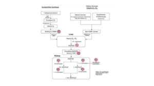
It’s what is laid out and implied in Fig. 1 above from the Deng, 2013 research. (Note all the red circles with magnesium!)

The next most important question to ask is why is the magnesium low in the Liver, in the first place?

I had originally completely overlooked this truth for the longest time up to this week.

It turns out that what I, and most who study on this topic, had successfully bypassed is, the fundamental fact that cholecalciferol (pro-Vitamin D3) is transformed in the liver by a key enzyme called hepatic 25-hydroxylase that we know is magnesium dependent. 

It is then subsequently transformed in the kidney by another key enzyme, renal 1a-Hydroxylase that is also magnesium dependent. We know all that.

What hit me like a ton of bricks was the realization that there is a reason why the liver doesn’t work right, and why it doesn’t allow proper function of that first key enzyme to work?

Anyone want to venture a guess as to why? 

It is none other than our rusty friend; excess, unbound iron being the proverbial “wrench in the works”. 

Chow, et al (1985) clearly state in their stunner of an article:

Chow, L.H., et al. (1985). “Low Serum 25-Hydroxyvitamin D in Hereditary Hemochromatosis: Relation to Iron Status.”
https://www.gastrojournal.org/article/S0016-5085(85)80001-9/pdf?fbclid=IwAR3Twk9RGQ-yP2xJMor7HTEjdbVhPanK29G74Pg8oRFtqt4Jl2at5MlubxU

“Under normal conditions, plasma 25(OH) D is formed in the liver from vitamin-D [cholecalciferol] absorbed from the diet or synthesized in the skin under the influence of ultraviolet light [8].” 

Emphasis on the word normal! They go on to point out that 

“This suggests that iron may have had a direct effect on serum 25(OH) D levels.”

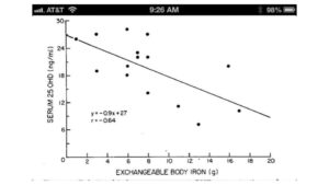
What is particularly poignant is the Fig. 1 that is from Chow et al, 1985; note the inverse correlation, as the iron level rises, the level of storage-D [25(OH) D] drops.

For those that think that this was a one-and-done study by Chow, et al; I happened upon an even more recent study by Ezzat, et al, 2015: (noted previously at the start of this post)

Ezzat, H.M., et al. (2015). “Vitamin D Deficiency and Liver Iron Concentration Transfusion Dependent Hemoglobinopathies in British Columbia”

https://pdfs.semanticscholar.org/410c/13eed8611f77310422696f0fcf143ae892e8.pdf

This study is a stunner, largely because it draws the same conclusion i.e. iron’s impact on storage-D levels (25 (OH) D) and has a notable set of Figures, as well. 

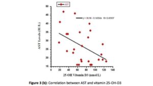
The most notable is the noted inverse relationship between a key liver enzyme, AST (sensitive to iron status), and level of Storage-D as noted in Fig. 3b:

Once again, the conclusion is inescapable; it is the iron getting in the way of the critical conversion of cholecalciferol in the liver.

Then it dawned on me, I knew why there is and has been such a rush to pump us up with synthetic hormone-D, that should have been naturally occurring in our liver, but could not. Not only that we lacked the magnesium; it is also that somewhere within the body system that we simply have too much iron in our livers to allow this essential metabolic conversion to take place.

Then I came across this research by Mangin, et al, 2014 takes on an even greater importance:

Mangin, M., et al. (2014). “Inflammation and vitamin D: the infection connection.”
https://www.ncbi.nlm.nih.gov/pmc/articles/PMC4160567/?fbclid=IwAR3IDqtDVchLgr8ISeQWhPLt49XwPbI7ZcoZzQuf81ON6DvgJinN0seYfiY

The connection between excess, unbound iron and the state of inflammation is absurdly high. The relationship between elevated ferritin, and inflammation is well documented. The connection between low magnesium and initiation of the entire inflammatory cascade, which I have repeatedly shared via the pioneering & penetrating research of Weglicki & Phillips of GWU Medial Center, 1992a, b, c & d.
https://www.researchgate.net/scientific-contributions/39152975_W_B_Weglicki

So suddenly, we have a new model to work with,
low storage-D (25(OH) D) levels are showing a billboard for both: 

“Too much iron and too little magnesium in the liver that together cause inflammation.”

Wow, that’s a stunner. It gives greater credence to the increasingly recognized condition of “Anemia of Chronic Inflammation”.

What is this research about iron <> vitamin-D is seeking to tell us?

Low storage vitamin-D is both a sign for low magnesium, and excess, unbound iron. Low storage-D (25(OH) D) is not a metabolic state needing to be corrected. It is signaling the very minerals (low magnesium and unbound excess iron) that need to be corrected!

Intriguing?

To bring this treatise to a noteworthy close, it is well known that James A. Holick, PhD, MD is the clinician that discovered that calcifediol (storage D – 25(OH) D) is the predominant form of Vitamin-D (hormone D) in the body, that calcitriol (active D – 1,25-(OH2) D) is the active form of vitamin D (hormone D). 

Holick was also the one that discovered that hepatic 25-hydroxylase is the very enzyme (noted earlier) that converts cholecalciferol >> calcifediol in the liver. He is the man when it comes to knowing all there is to know about hormone-D.
https://www.bumc.bu.edu/busm/profile/michael-holick/

But what I find fascinating is that not one reference can be found with his name and either of these key minerals of magnesium, or iron. Call me an odd duck, but I am deeply troubled by that scientific gap in his research, esp. when so many are being drowned, worldwide, by this synthetic, toxic form of vitamin-D, also known as “19-nor” in the research literature. I’ve no idea what that “19-nor” is referring to, but I fully intend to find out.

So, there you have it. We’ve compiled quite the year’s worth of new information by putting the spotlight on the dynamics of copper<>iron dysregulation and the related impact these two minerals then have on our magnesium status, in particular, as well as our physiology, overall. I plan to do an overview post on Hormone-D in the New Year, that I’m confident many of you will find both illuminating and reassuring.

Have a blessed New Year’s celebration later this week, and let’s look forward to new mineral insights and understandings in 2017!

A votre sante!
Morley M. Robbins

For Facebook Discussion:
www.facebook.com/groups/MagnesiumAdvocacy/permalink/1217780874956646/

Iron Toxicity Post #49: IP-6 offers a compelling solution to iron overload

Iron Toxicity Post #49: IP-6 offers a compelling solution to iron overload

IP-6 offers a compelling solution to iron overload.

We’ve covered a lot of ground this year as we’ve exposed the truth about the downside to excess, unbound Iron. I never imagined that I would spend this entire year (and likely the rest of my life) being consumed by this iron topic. We just never know what life has in store for us, now do we?

I am often asked, both on MAG, and consults, as to what can be done to lower the levels of iron in one’s blood. The most obvious action is to donate blood. Only 5% of folks regularly engage in this practice, but that small percentage of society outlives the other 95% in every study done to assess factors influencing longevity.

A close second to this phlebotomy practice is to discipline your supplement routine to take IP-6 (Inositol 6-Phosphate) or otherwise known as stabilized rice bran. The 6 phosphate groups (PO3), shown in the picture below, can hold an atom of iron which is a good thing in a body that is stressing over too much unbound iron.

I am sharing important information about this topic to two authors that I respect and both of who have compiled compelling blogs on this vital topic:

Bill Sardi on the “Overlooked Cancer Cure”:
https://www.lewrockwell.com/2005/06/bill-sardi/the-overlooked-cancer-cure-from-japan/

Joe Cohen on “16 Benefits of Phytic Acid

Between these two blogs, I’m confident that you’re getting a very complete overview, as well as drill-down, about the properties, the importance, as well as the impact that this anti-nutrient provides. Especially so, when it is taken away from food.

Here is a bit more scientific proof beyond what was provided in the those two blogs, please go below for additional research and analytical insights:

Weglarz, L., et al. (2008). “Effect of Inositol Hexaphosphate on Lipopolysaccharide-Stimulated Release of TNF-α from Human Mononuclear Cells”
https://pdfs.semanticscholar.org/0b26/00833995ff38def1db097d0332ff5d58302f.pdf

“It is thought that anti-carcinogenic benefits of IP6 may in part be attributable to its antioxidant capability through its iron chelating properties.”

Human mononuclear cell

Sandberg, A., et al. (1999). “Inositol phosphates with different numbers of phosphate groups influence iron absorption in humans”
ajcn.nutrition.org/content/70/2/240.full.pdf

“Inositol hexaphosphate (IP6) is a well-known inhibitor of iron absorption…”

Anekonda, T.S., et al. (2011). “Phytic acid as a potential treatment for Alzheimer’s pathology: evidence from animal and in vitro models”
www.ncbi.nlm.nih.gov/pmc/articles/PMC3021000/pdf/nihms234956.pdf

I believe when the push comes to shove, this nutrient is a powerhouse that will change your cellular chemistry. I do hope you find this information helpful and healthful.

A votre sante!
MORLEY M. ROBBINS

For Facebook Discussion:
https://www.facebook.com/groups/MagnesiumAdvocacy/permalink/1213843282017072/

Iron Toxicity Post #48: Dealing with cancer

Iron Toxicity Post #48: Dealing with cancer

This post is for about dealing with cancer.  I am going to be quite blunt and say that cancer is not a medical disease.  Iron causes cancer. If you still have serious doubts about this obvious metabolic connection please take a few moments to read this momentous study:

Tesfay, L., et al. (2015). “Hepcidin Regulation in Prostate and Its Disruption In Prostate Cancer.”
https://cancerres.aacrjournals.org/content/canres/75/11/2254.full.pdf

Keep in mind the authors go to great lengths to suggest that this only applies to prostate cancer, which is the #1 cause of cancer in men.  It is also the second leading cause of cancer-related deaths as well. 

This study has changed how I look at how this works and I disagree with the narrowcasting of these results. As you may know from earlier posts, my Dad died when I was 16 of lung cancer, then my Mom’s older sister died of liver cancer and my sister has had double breast cancer and lives to see another day means that I am no stranger to this tragic condition.

I believe the points made in this study, i.e. the role of iron in promoting cancer are universal and should totally change our perception of what actually causes “cancer”.

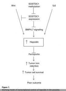
I have provided a dot connecting points as I synthesize this research:

  • Inflammation >> 
  • Presence of cytokines (IL-6) >> 
  • Increased production of hepcidin >> 
  • Increased destruction of ferroportin >> 
  • Increased retention of iron in the cancer cells >> 
  • Increased carcinogenic activity of the cell.

I want to draw your attention to Fig. 7. noted below that highlights this cascade of events with particular emphasis on the increase of hepcidin that blunts ferroportin’s key function to ensure proper iron circulation.

This blunting of ferroportin then leads to three stunning outcomes in those with cancer:

  • Increased retention of iron within the tumor 
  • Increased tumor cell survival 
  • Increased likelihood of a poor clinical outcome 

As you know from my previous iron toxicity posts about iron and copper metabolism, this study reminds you that iron is regarded as the perfect carcinogen.

Did you know that it is the only element on this planet that meets all three criteria to be considered a carcinogen?

  1. Iron initiates cancer
  2. Iron promotes cancer
  3. Iron progresses cancer

Source: Moon, J. (2008). “Iron: The Most Toxic Metal.”

Dr. Moon goes on to describe the unique characteristics of this ultimate carcinogen by noting, and I quote from his mind-bending book:

  • Iron activates xenobiotics (foreign chemicals) turning them into carcinogens
  • Iron is a factor leading to alcoholic liver cirrhosis, a precursor to liver cancer
  • Iron acts synergistically with viral infections in causing cancer
  • Iron multiplies aflatoxin (hepatic mycotoxin from mold) carcinogenicity
  • Iron plays a critical role in causing cancer cells to become more aggressive and invasive

This pretty much seals the deal on how and why cancer rates have risen to outrageous levels through this iron dominated planet. 100 years ago, the incidence of cancer was 1 in 20. Today it is 1 in 2. 

The cause of cancer is iron. There is some confusion about the origin of inflammation but I encourage you to look past what it is thought of and follow the compelling dots points of how this same magnesium deficiency and iron toxicity dynamic affects a mineral deficient body. Not to mention how it creates all manner of chronic conditions such as cancer:

  • Inflammation is caused by magnesium deficiency 

Weglicki, W.B., Phillips, T.M. (1992). “Pathobiology of magnesium deficiency: a cytokine/neurogenic inflammation hypothesis.”
https://www.physiology.org/doi/abs/10.1152/ajpregu.1992.263.3.r734

Inflammation is not a medical disease. It is, however, a clinical sign of magnesium deficiency and initiates the production of substance P, which triggers the inflammatory cascade of all cytokines and chemokines. NF-kB initiates the release of IL-1a, IL-6, and many other peptides. Note that this is the origin of the IL-6 that plays such a key role to start the synthesis of hepcidin in the prostate tissue. I would argue, this is happening in tissue all over the body.

  • It is a known fact that iron accumulates in the tissue of magnesium deficient critters (4-legged & 2-legged!):

Kimura, M., Yokoi, K. (1996). “Iron accumulation in tissues of magnesium-deficient rats with dietary iron overload.”
https://www.ncbi.nlm.nih.gov/pubmed/8907021

Sanchez-Morito, N., et al. (2000). “Influence of magnesium deficiency on the bioavailability and tissue distribution of iron in the rat.”
https://www.semanticscholar.org/paper/Influence-of-magnesium-deficiency-on-the-and-tissue-Sanchez-Morito-Planells/4a9799be01dbae10ae3f003c8f3bc0d52a02b298

I think it is highly relevant that in the former study, they note that 

“…magnesium deficient rats with iron overload may be used as an experimental hemochromatosis model.”

  • It appears that hepcidin expression is increased in critters that are magnesium deficient:

 Ishizaki, N., et al. (2011). “Hepcidin expression in the liver of rats fed a magnesium deficient diet.”
https://www.ncbi.nlm.nih.gov/pubmed/21736832

Note many of the same players, especially BMP-6, are described in this process of increased iron accumulation in the tissue of a magnesium deficient body. I believe that most critters on this planet are magnesium deficient! Therefore, most stand the increased chance for increased iron storage.

 Now the literature is relatively silent on what I believe is a metabolic fact: Iron causes Inflammation. So, given my inspired assertion:

  • If magnesium deficiency causes inflammation
  • Iron builds
  • Hepcidin expresses more in magnesium deficient tissue

Do you think that iron is causing inflammation? This then leads to a slight modification of the initial dot connecting:

Iron causes inflammation >>> 

Presence of cytokines (IL-6) >>> 

Increased production of hepcidin >>> 

Increased destruction of ferroportin >>>

Increased retention of iron in the cancer cells >>>

Increased carcinogenic activity of the cell

The build up of iron due to the systemic lack of magnesium and ceruloplasmin causes the very inflammation that triggers the cellular signaling toward carcinogenic activity that then causes further iron retention leading to further cancer activity. 

The circular nature of this destructive pathway is mind numbing. What’s the one word that is totally missing in this scintillating study on prostate cancer? Ceruloplasmin!

Here is the gist of why we are all in such a quandary:

“Therefore, the ceruloplasmin-ferroportin system represents the main pathway for cellular iron egress and it is responsible for physiological regulation of cellular iron levels.” 

Musci, G. et al. (2014). “Ceruloplasmin-ferroportin system of iron traffic in vertebrates.”
https://www.ncbi.nlm.nih.gov/pmc/articles/PMC4050113/pdf/wjbc-5-204.pdf

The iron-based literature goes to great lengths to create a connection between hepcidin <> ferroportin, but they totally ignore ceruloplasmin. Keep in mind, Hepcidin, is a hormone with 25 Amino Acids. Ceruloplasmin (Cp), on the other hand, is one of the most important enzymes to grace the human body, has 1,046 amino acids and 8 copper atoms (4 Cu+/4 Cu++ surrounding an oxygen molecule). Yes, we can all certainly understand why they would ignore this powerhouse metabolic player, given the dynamics of clinical actions today.

I would encourage you ALL to read this critical study by Musci, et al., to better understand how to keep hepcidin from going rogue and thus causing a build-up of stored iron in your tissue that will eventually lead to a carcinogenic growth.

Tesfay, et al., 2015, the first article I mentioned completely overlooked this foundational enzyme fact in their study on prostate cancer should send a chill down our collective spines. How do they not know or even acknowledge the known regulatory role of ceruloplasmin and its critical impact on the regulation of the management of iron inside and about the cell?

I implore you to realize how the hallowed halls of clinical research that these iron-focused researchers are doing, connecting critical dots and at the same time overlooking pivotal pieces of the puzzle. 

A votre sante!
MORLEY M. ROBBINS

For Facebook Discussion:
www.facebook.com/groups/MagnesiumAdvocacy/permalink/1211975545537179/