Australia – the Root Cause Protocol World Tour Begins.

Australia – the Root Cause Protocol World Tour Begins.

Cold weather, snow, ice and short daylight days getting you down?

Here’s a “prescription”!
Go on an education to Australia!

That’s right, it’s summer in the Southern Hemisphere and Morley Robbins and Dr.Liz will be there conducting consults, workshops and Root Cause Protocol Consultant training leading to certification in Melbourne, Sydney, Brisbane and Perth.

What can be more ideal?
Don’t wait, as February 2018 comes soon. To learn more…. the details, four locations, registrations and etc., go to:
The RCP World Tour in Melbourne, Sydney, Brisbane and Perth, Australia.
http://www.rcpworldtour.com
See you Down Under in February!
Morley Robbins

P.S.   If you can’t get away, here are two options:

1.  learn about the Root Cause Protocol via this new video series.
http://therootcauseprotocol.com
2.  to become a Root Cause Protocol certified consultant, the next 16 week online intensive training begins March 8, 2018.  Register now to secure your place.
http://gotmag.org/train-with-us/

Iron Toxicity Post #66: ‘Iron and the RBC (Red Blood Cell): Is this where the assault begin?

Iron Toxicity Post #66: ‘Iron and the RBC (Red Blood Cell): Is this where the assault begin?

“Iron and the Red Blood Cell (RBC): Is this where the assault begins?” (OMg! Does a bear sleep in the woods?) 

Dr Liz and I just watched the new movie on P.T. Barnum: “The Greatest Showman.” It’s definitely worth your time to witness the captivating story behind the indomitable spirit and imagination of this amazing figure in our entertainment past. The movie ended with one of the most famous quotes attributed to him:

“The noblest art is that of making others happy.”

I really like that sentiment, but I’m going to go one step further, and paraphrase it:

“The noblest act is teaching others how to be healthy.” 

(I intend to adopt that statement as my signature effort in this lifetime)

I am devoting the rest of my days to helping people on this planet to get educated and to get empowered on the key steps that need to be undertaken to re-store metabolic balance in their bodies. It is not rocket science!

I should add that I am hardly alone in this effort, and among my many “mentors” that have helped shape and mold my thinking about these key requirements is Jerry Tennant, MD (Dallas, Texas), recently praised with a Lifetime Achievement Award at the Doctors Who Rock symposium in Florida. He is also the noted author of “Healing is Voltage,” among other books, which is a must read for anyone wanting to understand the wiring of the human metabolism, and the vital importance of cellular energy production to assure optimal cellular frequency therefore optimal health.

It’s worth knowing that what inspired Jerry Tennant, MD, at the depths of his 7-yr long illness crisis, was the fervent belief that if he could get one cell in his body to work, then he could get them all to work! (I should add, this focus is akin to the thinking of a gifted mechanic that I worked with, named Otis, one summer while on break from college. He was convinced that if he could get one cylinder in the engine to work, he could get them all to work.)

Given that focus, what then do I regard as the most important cell in the human body?

Well, I would argue that it’s the erythrocyte, as known as the red blood cell, or RBC. It’s been on my radar screen for ~8 years!

Okay, here are some key facts that highlight the importance of this ubiquitous and largely overlooked cell in our body:

  • There are some 20-30 trillion RBCs in the average adult
  • In one small drop of blood (1 cubic mm) there are 4-5 million RBCs
  • RBCs account for ~84% of all human cells. (Please let that % sinks in before dashing to the next line.)
  • RBCs, by virtue of being created inside the bone marrow, are considered connective tissue. (Again, please let the significance of that fact sink in!)
  • Each human RBC contains ~250-270 million hemoglobin molecules
  • There are 4 heme proteins contained in each molecule of hemoglobin, and each has an atom of iron in the center. (Not unlike a ceiling fan in the center of your den or living room)
  • Each human red blood cell can carry 1 billion oxygen (O2) molecules (And if iron is misbehaving, that is a lot oxygen molecules to create potential rust, folks)
  • 70 % of all iron contained in the human body is found in those zillions and zillions of haemoglobin
  • The oft-quoted ideal lifespan for these RBCs is ~120 days
  • The rate-limiting enzyme to determine the energy, viability and thus lifespan of these RBCs is G6PD enzyme, aka Glucose-6-Phosphate Dehydrogenase (It’s the head of the stream of the pentose phosphate pathway)
  • “G6PD deficiency” is regarded at the greatest enzyme deficiency on this planet. It’s worth noting that this enzyme has relationships with both magnesium and bioavailable copper.
  • When RBCs get stressed and can’t make proper energy, they lose their flexibility, and energy-driven shape, and then get “flagged” for re-cycling, as known as hemolysis.
  • In separate studies, either copper deficiency or magnesium deficiency causes the RBC lifespan to drop 80% to ~20 days! That is an extraordinary loss of lifespan, and an extraordinary acceleration of RBC turnover!
  • This turnover is the very metabolic mechanism that leads to iron-laden macrophages that results in all autoimmune conditions.
  • It is worth repeating that the synthesis of heme, the insertion of iron into heme, the knitting of four heme to make one hemoglobin molecule, as well as the regulation of HO-1 (heme oxygenase-1) “re-cycling” enzyme all require bioavailable copper.
  • Where does oxidative stress and inflammation start in our bodies? Right here in the RBCs (Red Blood Cells) of our blood.
  • Most importantly, please remember that iron simply carries oxygen throughout the body, but for a fact, copper is the only mineral that can use or activate oxygen to release energy or change chemicals (e.g. neurotransmitters) in the body. That is a major big deal that is lost in the world of conventional “you need more iron” medicine.

Given all of this, doesn’t it make sense to start the process of understanding how to stop the oxidative chaos that originates inside the red blood cells?

Call me goofy, but this seems to me like a penetrating look into the obvious. Thankfully, I am not alone in this thought, as it has been a major focus of research for many decades. Regrettably, none of these findings about the importance and the fragility of the RBC seems to be taught at any school of health practitioner training anywhere on the planet in this modern era.

Here are some noted studies that offer keen insights to this iron <> RBC foundational premise:

Clemens, M.R., Waller, (1987). “Lipid peroxidation in erythrocytes”
www.ncbi.nlm.nih.gov/pubmed/3319229

“Most of the protective activity [for RBCs] is associated with two proteins, transferrin and ceruloplasmin.” 

As has been noted before, iron requires constant supervision or chaperoning, not unlike a 4-yr old with a hammer!

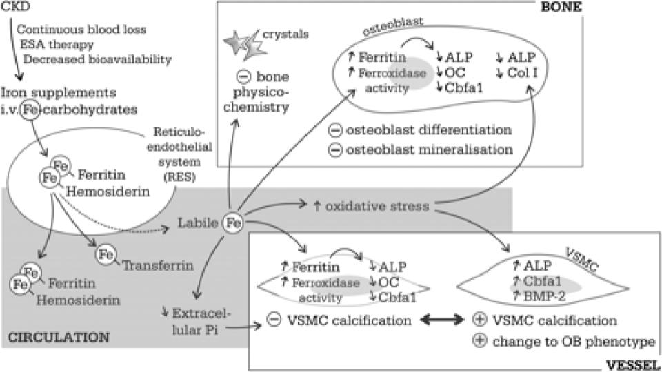
Neven, E., et al. (2011). “Iron and vascular calcification: Is there a link?”
academic.oup.com/ndt/article/26/4/1137/1884318

Figure 1 (below) of this seminal assessment of iron-induced calcification of patients with CKD is stunning. The fact that RBCs are considered connective tissue and have osteogenic [bone-making] properties is a game changer in terms of the recognized stimulatory role of excess, unbound iron in triggering and fostering plaque (atherosclerosis) and hardening of the arteries (arteriosclerosis), as well as calcification of organs throughout the body.

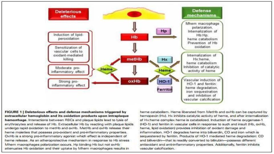
Jeney, V., et al. (2014). “Red blood cell, hemoglobin and heme in the progression of atherosclerosis”
www.ncbi.nlm.nih.gov/pmc/articles/PMC4183119/

“Neovascularization is induced mostly by hypoxia [lack of oxygen, but also lack the ability to use oxygen] but the involvement of oxidative stress is also established.” 

When the speed of hemolysis accelerates, the protective proteins to guard hemoglobin [haptoglobin] and heme [hemopexin] are simply used up!

Zarjou. A., et al. (2010). “Ferritin ferroxidase activity: A potent inhibitor of osteogenesis”
onlinelibrary.wiley.com/doi/10.1359/jbmr.091002/full

“… osteogenesis and pathogenesis of iron caused bone defects, and by exposing ferritin as the cause of decreased OB [osteoblast] maturation…” 

The excess of iron is the true bad boy in osteoporosis, and the role of ferritin is key, especially when it has ferroxidase enzyme function.

Vercellotti, G.M., et al. (2014). “H-ferritin ferroxidase induces cytoprotective pathways and inhibits microvascular stasis in transgenic sickle mice”
www.frontiersin.org/articles/10.3389/fphar.2014.00079/full

“We conclude that wt-hFHC (wild type-human ferritin heavy chain) ferroxidase activity enhances cytoprotective Nrf-2-regulated proteins, including HO-1, thereby resulting in decreased NF-kB activation [key marker for inflammation], adhesion molecules, and microvascular stasis in transgenic SCD [Sickle Cell Disease] mice.”

Once again, ferroxidase enzyme function protects via its anti-oxidant properties and functions throughout the body.
 
So, what’s the bottom line?

We are left LOST in the Byzantine world of allopathic blood testing. There are no tests for:

  • Ferroxidase enzyme function (Ceruloplasmin is a proxy, but not the real deal)
  • No distinction between ferritin- heavy chain vs. ferritin light chain, and no assessment of either in intracellular ferritin!
  • Oxidized low-density lipid (LDL) cholesterol, a key marker for oxidative stress in the blood.
  • Neither haptoglobin, nor hemopexin are measured, which are key to protecting hemoglobin and heme, respectively in the blood, as noted above.
  • No marker for hemosiderin, otherwise known as de-natured ferritin from too much hydrogen peroxide (H2O2).
  • No marker for RBC lifespan to better understand the rate of hemolysis (RBC turnover)
  • No marker for hydrogen peroxide (H2O2), which is a major oxidant to cause RBC chaos.
  • No easily accessed marker for assessing iron status inside the red blood cell, where it matters most.
  • No easily accessed marker for erythrocyte superoxide dismutase (SOD), the most sensitive marker for the bioavailability of copper in the human body.

 Trust me, I could go on, and on with the limitations of conventional iron testing!

I think you get the point at this point in the process. The healthier our red blood cells (RBCs) are, the healthier we are. The potential for oxidative assault in our blood that is largely overlooked is quite significant. That is why we obsess over the level of magnesium in the RBC. Isn’t it odd that no doctor that you’ve ever been to has ever tested for this seminal mineral inside this seminal blood cell? Personally, I find that fact alone, staggering and incriminating. In light of the research shared above, you should too!

The better regulated by copper, and the better chaperoned by proteins our iron is, the less likely we are to be subjected to oxidative stress and/or inflammation. The research is quite clear about that, following four years of intensive study on that one dynamic alone!

So, the Root Cause Protocol has particular relevance and reason for its health-enhancing impact. 

I will leave you with one last quotation from P.T. Barnum:

“The foundation of success is good health.”

He was the consummate showman, but I would offer that he was also a great “Sha-man”!

A votre sante!
Morley M. Robbins

For Facebook Discussion:
https://www.facebook.com/groups/MagnesiumAdvocacy/permalink/1570321223035941/


Iron Toxicity Post #65: There is NO such thing as ‘Iron Metabolism’.  There is ONLY CopperIron Metabolism

Iron Toxicity Post #65: There is NO such thing as ‘Iron Metabolism’. There is ONLY Copper<>Iron Metabolism

How does iron regulate itself?

I’m sharing the musings of just one day of my efforts to research iron, and what’s posted below is only ½ of what I found. I am forever amazed at the lengths that iron researchers will go to slice and dice their findings and insights, but overlook the most important fact.

In these nine articles and there are 100’s more just like them that examine the many facets of iron metabolism and iron homeostasis, there is one word that is missing from all of these studies. Any one want to venture a guess as to what that one word might be?

Yes, that word would be copper!

They do make a point of mentioning ceruloplasmin, and on a rare occasion, will discuss the ferroxidase enzyme function, but no where do they honor or highlight that there is no such thing as iron metabolism.

There is only copper<>iron metabolism, and as I’ve noted many times, iron is, for a fact, the dummy in that metallic tango!  Despite what you read on the Internet, please trust me, iron takes its cues from bioavailable copper. Solely!

And what is most important to understand is that iron is not designed to be in storage, 80% of the iron in your body is found in hemoglobin and myoglobin, which is in constant circulation. <10% is found in ferritin and that is meant to be inside the cell, not inside your serum!
The study by Vanoaica (2010, below) is unique in its recognition that ferritin-H is essential for accurate control of iron. What is ferritin-H (Heavy) chain?

It is a form of ferritin that has ferroxidase enzyme function, which is essential for proper iron egress and circulation. (They have known this since 1968!)

Before you get too excited, no, your doctor has no way to distinguish this form of ferritin from the ferritin-L (Light) chain. You would think that they would, but alas, we are left with the mushrooms, in the dark and buried in compost! In your next life you’ll want to come back as a 4-legged rat, so that you’ll know for certain what your ferritin fractions are, as these two tests are done all the time in the research labs.

  1. Chen, C., Paw, B.H. (2012). “Cellular and Mitochondrial Iron Homeostasis in Vertebrates.”
    https://www.sciencedirect.com/science/article/pii/S0167488912000055
  2. Anderson, C.P., et al. (2012). “Mammalian iron metabolism and its control by iron regulatory proteins”
    doi.org/10.1016/j.bbamcr.2012.05.010 
  3. Hentze, M.W., et al. (2010). “Two to Tango: Regulation of Mammalian Iron Metabolism”
    doi.org/10.1016/j.cell.2010.06.028
  4. Kuhn, L.C. (2009). “How Iron Controls Iron” Cell Metabolism 10(6): 439-441
    https://www.sciencedirect.com/science/article/pii/S1550413109003428
  5. Ganz T. (2008). “Iron Homeostasis: Fitting the Puzzle Pieces Together” Cell Metabolism 7(4): 288-90
    https://www.cell.com/cell-metabolism/fulltext/S1550-4131(08)00079-X?_returnURL=https%3A%2F%2Flinkinghub.elsevier.com%2Fretrieve%2Fpii%2FS155041310800079X%3Fshowall%3Dtrue
  6. Simpson, R.J., et al. (2009). “Regulation of Intestinal Iron Absorption: The Mucosa Takes Control?” Cell Metabolism 10(2):84-87
    https://www.cell.com/cell-metabolism/fulltext/S1550-4131(09)00189-2?_returnURL=https%3A%2F%2Flinkinghub.elsevier.com%2Fretrieve%2Fpii%2FS1550413109001892%3Fshowall%3Dtrue
  7. Andrews, N.C. (2010). “Ferrit(in)ing Out New Mechanisms in Iron Homeostasis” Cell Metabolism 12(3)-203-204
    https://www.cell.com/cell-metabolism/fulltext/S1550-4131(10)00279-2?_returnURL=https%3A%2F%2Flinkinghub.elsevier.com%2Fretrieve%2Fpii%2FS1550413110002792%3Fshowall%3Dtrue
  8. Vanoaica, L., et al. (2010). “Intestinal Ferritin H Is Required for an Accurate Control of Iron Absorption” Cell Metabolism 12(3):272-283
    https://www.cell.com/cell-metabolism/fulltext/S1550-4131(10)00268-8?_returnURL=https%3A%2F%2Flinkinghub.elsevier.com%2Fretrieve%2Fpii%2FS1550413110002688%3Fshowall%3Dtrue
  9. Thompson, J.W., et al. (2012). “Protein degradation and iron homeostasis” Biochimica & Biophysica Acta (BBA) – Molecular Cell Research 1823(9):1484-1490
    https://www.sciencedirect.com/science/article/pii/S0167488912000328?via%3Dihub

And what these many studies are seeking to reveal is that the regulation of Iron relies on two key pathways for optimal iron circulation and R.E.cycling:

  • Ferroportin<>hepcidin system, but they are silent on the fact that this system does not work without bioavailable copper in the form of ferroxidase enzyme function.
  • Iron regulatory proteins (IRP1-2) and iron response elements (IREs) System, but they are silent on the fact that this system responds equally well to retinol (animal-based Vitamin-A) just as well as is does to iron supplementation. What is fascinating about that factoid is that retinol is key and foundational for the synthesis of ferroxidase enzyme need above. 

C’mon now, don’t you find that just a tad bit entertaining

Folks…

We are drowning in hormone-D supplements that kill retinol status in the liver!

We are drowning in iron supplements and infusions that kill our metabolism!

We are drowning in zinc supplements that cause copper to be bound up, and be not available.

We are drowning in ascorbic acid supplements that increase H2O2 (which tweaks the ceruloplasmin protein and kills the ferroxidase function) and accelerate iron storage.

Am I the only one seeing a predictable pattern here with the nutritional practices of allopathic and alternative practitioners?

I wish it weren’t this obvious, this clear and just wait until I share the other ½ of today’s research that states definitively that cancer is caused by iron. I almost fell out of my chair earlier today!

Better MAG up, and be prepared for the next iron toxicity post!

A votre sante!
Morley M. Robbins

For Facebook Discussion:
https://www.facebook.com/groups/MagnesiumAdvocacy/permalink/1543368645731199/

Iron Toxicity Post #64: Ferroxidase blocks the Hydroxyl Radical (OH*)

Iron Toxicity Post #64: Ferroxidase blocks the Hydroxyl Radical (OH*)

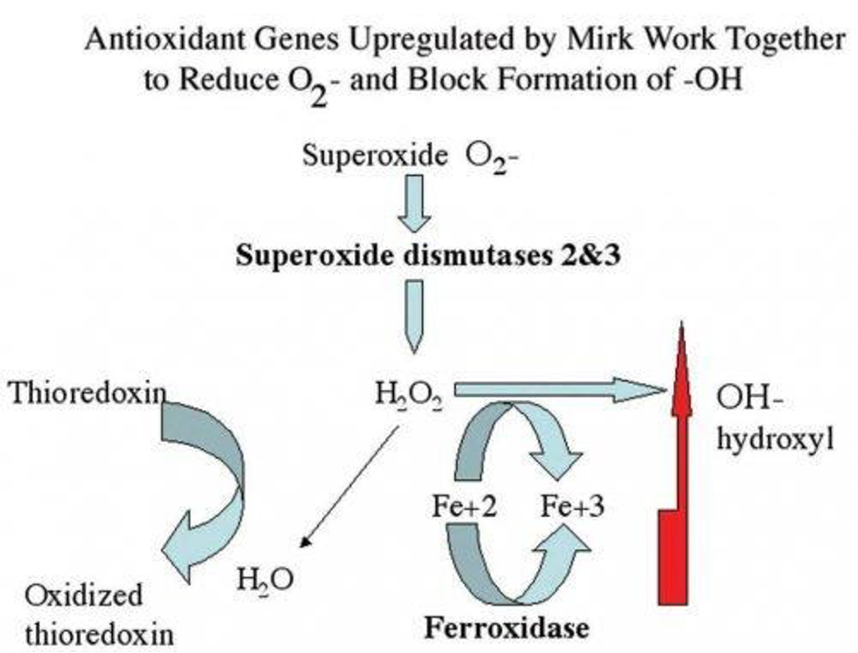
Ferroxidase blocks the hydroxyl radical (OH*).

I am speechless to learn this truth.

I learned it here at Fig 8:
www.mdpi.com/2072-6694/2/3/1492/htm

I will say more tomorrow about the foundational importance of this aspect of the ferroxidase enzyme and why the root cause protocol is so important!

View my new video series:
https://therootcauseprotocol.com/rcp-101-video-series/ 

A votre sante!
Morley M. Robbins

For Facebook Discussion:
https://www.facebook.com/groups/MagnesiumAdvocacy/permalink/1501091929958871/

Iron Toxicity Post #63: Setting the story straight about iron homeostasis

Iron Toxicity Post #63: Setting the story straight about iron homeostasis

Setting the record straight about iron homeostasis.

The critical dynamic that is kept from us is that iron homeostasis is a seesaw that has two sides to it, as well as a fulcrum in the middle:

  • One side = iron in the blood
  • Fulcrum = ferroxidase enzyme function (as know as, FOX, Cp and ceruloplasmin)
  • Other side = iron in the tissues

The part that we missed is that just like any seesaw, when one side is heavy, the other side will be sky high!!! Get it?

Anemic (not enough iron in the blood) really means that it’s lots of iron in the tissue! 

Iron is like a square dance, it is meant to be in constant circulation, just as any good square dance you have ever been in or seen. The emphasis is on motion.

Iron has become akin to a motion picture, which involves no motion on the part of the iron (observer!), as now the clinical focus is on being stored, as reported in the ferritin storage protein!

That is not a natural state for iron, and not as originally intended, please understand that stored iron leads to rust!

The purpose of the Root Cause Protocol is to provide the set of nutrients to re-engage the square dance and get the iron out of those seats of the movie theatre.

Here’s the ugly part: the yardstick to properly measure iron homeostasis currently does not exist!

  • The “One side” measures ferritin, which is a flawed marker that is found inside the cell, and when ferritin is outside of the cell, it indicates liver pathology and copper dysregulation.
  • The “Fulcrum” is not properly measured as the commercial labs are instructed to not measure the ferroxidase enzyme function and just report “generic” ceruloplasmin, which is an insult to our intelligence and our integrity.
  • The “Other side” is never measured easily as the only options are a needle biopsy (very painful!) or a r2/t2 MRI (very expensive!). 

We simply have no valid or meaningful measure of iron saturation in the tissues, where the other half of the iron story needs to be told!

So, until the metrics get into the 20th Century, please do the Root Cause Protocol, and know that your ongoing process to rebuild cells and properly re-circulate the iron will enable a “reversal” of the stuck iron causing the neurodegeneration because no one ever told you how iron was really supposed to work inside our blood and inside our tissue.

A votre sante!
Morley M Robbins

For Facebook Discussion:
https://www.facebook.com/groups/MagnesiumAdvocacy/permalink/1474110502657014/