New RCP Website – 2018

New RCP Website – 2018

Even a mighty oak tree was once a tiny acorn…

It is with great excitement (and relief) that I announce the re-launch of this website, TheRootCauseProtocol.com

Previously, this website only hosted the RCP 101 video series. (And if you’ve previously purchased the RCP 101 video series, you would have a received a seperate email with your new login details – please check your junk/spam folder)

Now, this website is THE HUB for all things RCP, including:

We have big plans for 2019 and beyond, including:

  • An RCP Podcast
  • More RCP World Tour events (the Detroit event was a blast!)
  • Much more…

A vôtre santé​,
Morley Robbins 

Thank you Patrons – RCP Pioneers!

Thank you Patrons – RCP Pioneers!

In May 2018, I launched a simple Patreon page for RCP.

Here’s what I posted in the very first Patreon message:

Facebook has been an amazing platform for the growth of MAG. But Facebook is sort of like a beach, and every new Post is like writing a message in the sand… It’s only so long before the tide comes up and it’s “lost” forever.

So I intend to use Patreon as a publishing tool (writing, videos, podcasts) for messages that need a bit more permanence. You, faithful supporters, will have exclusive access to this content.

In five short months, 287 patrons had joined Patreon to support these efforts.

Patreon was basically the prototype for the community I was hoping we would build some day on our own website.

Well, that day is here, and much sooner than I expected!  (Learn all about the new website here)

If you are Patreon monthly supporter, here’s what you need to do:

  1. Cancel your monthly Patreon donation as that website will be discontinued on November 30, 2018. (Don’t worry, all important content from Patreon has been migrated into the new Musings by Morley forum.)
  2. Join the new Premium RCP Community. ($9.97/month or $99/year for the first 2,000 “Foundation” supporters.)

So even though we’re moving on from Patreon, I still want to honor and memorialize all of the following “RCP Pioneers” (those who donated $5/month or more) for being the first to join up and support these efforts.  Thank you!

  • Amanda Bryce
  • Amy Armstrong
  • Andrea Macqueen
  • Ann Marie Case
  • Arne Kaminsky
  • Barbara J Alger
  • beth hardej
  • Betsy Kantor
  • Camilla Holing Eide
  • Carmen Wiles
  • Cathy Streitwieser
  • Christine Alexander
  • Colette van Haaren
  • Daniel Cooney
  • Daniela Canepa
  • Daniela Sola
  • Diane Beach
  • Doris Atkinson
  • Dr Catherine Julia Mort
  • Elaine Nikula
  • Elise Ellicott
  • Elizabeth Henderson
  • Florentina Marcu
  • Gine Elisabeth Høeg
  • Greta Hyatt
  • Heather Crimson
  • Heidi Harbaugh
  • Henrietta Bruun
  • Henry Townsend
  • Hope Godbee
  • Igor Meštrić
  • Ines Mitrovic
  • Ineta Kirse-Tiesniece
  • Inger Bletcher
  • Jacqui Soliman
  • Jane Clark
  • Jane martin
  • Jannie Sperling
  • Jenna Nowland
  • Jenny Ren
  • Jill Morris
  • Jill Peterson-Walton
  • Jodi Briggs
  • Johan Nilsson
  • John D Matthews
  • Julie Buckley
  • Karen Gay
  • kat farovitch
  • Kathleen Roussel
  • Kelly Harris
  • Kurt Geoffrey Staples
  • Laura Boerman
  • Laura Vida
  • Laura W
  • Lorinda Moore
  • Lynn Galbato
  • Lynn Morrow
  • madonna (susan) ferguson
  • Maggie Hucko
  • Marcia Allard
  • Margaret Peterson
  • Maria Townsend
  • Marta Tompkins
  • Mary Sue Powers
  • Mary Theresa Jurnack
  • Mary Wolfe
  • Matthew McCarthy
  • Melissa Kubala
  • Merav Srur
  • Meredith Moore
  • Michael Leone
  • Michele Norrid
  • Modesto Barra
  • Norma Edwards
  • Pat Scherler
  • Patrick Couchman
  • Patty Downs
  • Renay Ingledew
  • Rhonda Evans
  • Ricki Lindsay
  • Shalimar Little
  • Sheila Gardiner
  • Sheryl Winton
  • Stephanie Roberts
  • Susan Gagliano Coe
  • Teresa Bowley
  • Teri Linneman
  • Toni Little
  • Toril Bentzen
  • Tracy Tredoux
  • Twila J Starkey
  • ursula anderson
  • Valerie Engh
  • vasudha Tekriwal
  • Victoria Thomas
  • Virginnia Watt
  • Vivienne Howard
  • Wanda
  • Yvonne
  • Yvonne Boecker
  • Maree Perkins
  • Berit Lilja Staib
  • Brenda Rustick
  • Dallas Goodale
  • Dan Corrigan
  • David Krizo
  • Dominique Jones
  • Frank Lister
  • Helen Witts
  • Karen Brazeau
  • Kelly Veenstra
  • Laurel Wilkinson
  • M J H
  • Maria Guidry
  • Maria Montero
  • Maureen Clark
  • Nicola
  • Samantha Rodriguez
  • Shannon Tefft-Janes
  • Susan Plum
  • Tone Søndmør
  • Amanda Jefferson
  • Kang Kok Hua
  • Liv Irene Halvorsen
  • Christina Bell
  • Everett (Tim) Marvin
  • Irene Askildsen Andersen
  • Jack U
  • Jen Fokkens
  • Jens Veiersted
  • Karen Lemacks Gadol
  • Kay Moscinski
  • Laura Clegg
  • Linda Grytten
  • Marc Woods
  • Margie Yemini
  • Mariola Siwik
  • Robert Glantz
  • Robert Thompson
  • Judene Benoit
Iron Toxicity Post #75: PROOF that Copper Deficiency is the CAUSE of so-called ‘anemia’

Iron Toxicity Post #75: PROOF that Copper Deficiency is the CAUSE of so-called ‘anemia’

(Formerly #76)

One of the cardinal points of interaction and interface between copper <> iron is that the proper absorption of iron in the duodenum of our gut requires heme iron and/or reduced ferric iron (found in non-meat food sources) to enter the enterocyte via a key protein pathway called DMT-1, as known as Divalent Metal Transporter-1. That’s the front door into the 1st tissue that receives the iron in our gut, aka the enterocytes. But it’s the back door where the real action seems to take place, especially as it relates to iron.

At that other end, the enterocytes back up to the bloodstream, and for iron to get out of the enterocytes, and into the bloodstream, there are three critical factors to contend with:

  • Another doorway, called Ferroportin, aka FPN.
  • A doorman, called Hephaestin, (HEPH) which is a copper-dependent, ferroxidase enzyme that transforms ferrous (Fe2+) iron into ferric (Fe3+) iron so it can bind to transferrin; and
  • A regulator, called Hepcidin, (HPN) which is called the Iron Regulatory Hormone, but it’s also called the “Inflammation Hormone!” as Hepcidin’s production is triggered by the presence of either iron and/or a set of inflammatory cytokines, IL-1, IL-6 or TNF-a.

And when hepcidin shows up consistently, this peptide causes the degradation of that back doorway and the iron egress gets stopped, thus leading to the build-up of iron in the gut. Please know this process of inflammation-driven iron retention is what leads to many gut issues, like Leaky Gut, SIBO, Crohn’s, Colitis, etc.

These mechanisms to “catch & release” iron are the classical understanding of how iron “gets into the gut,” and either it “gets out” via the presence of copper-driven hephaestin, or it “gets stuck” via the increased prevalence of iron, and thus hepcidin, in the 1st line of tissues in our digestive system.

And all is good with this ^^^^ description above, but it overlooks a glaring issue: So if copper is needed to enable iron to get out of the gut, then how in the world does iron build up in tissue, all over the body if we’re all lacking bioavailable copper?

It’s a really important question and one of my Copernican students in Group #5 asked me this very question just the other day.

My initial reaction to this sincere question was one of frustration, as expressed by my initial response: “We don’t need to know that, just accept that the process of iron build-up happens…” Hardly a proper nor a complete response, and thankfully, her question just kept grinding away at me. I couldn’t explain with certainty how this iron dysregulation and subsequent accumulation was actually happening outside the gut, given that the iron was obviously not stuck there, even though copper is likely in a deficit position.

Then, by the grace of God, I found the following breakthrough study:

Matak, P. et al. (2013). “Copper Deficiency Leads to Anemia, Duodenal Hypoxia, Upregulation of HIF-2a and Altered Expression of Iron Absorption Genes in Mice”
https://www.ncbi.nlm.nih.gov/pmc/articles/PMC3610650/pdf/pone.0059538.pdf

Presented at the 1st Figure (Labeled “Fig 5” at the top of this post) is the key dynamic. What we see there as you follow the diagram from the bottom up and over to FPN (on the right side) is that in the presence of a copper-deficient organism, in this case rodents, is that copper deficiency leads to hypoxia – a profound discovery, which then affects the function of a key protein called HIF-2a, as known as Hypoxia Inducible Factor-2alpha, which is a powerful protein that responds to the “lack of oxygen” signal, and triggers the opening of that key FPN doorway to allow iron out of the enterocyte and into the bloodstream.

Before we go any further, a quick digression. “Hypoxia,” as it turns out, is a double-entendre:

  • “Low” oxygen implies low levels of iron, given that iron is key to “carrying” oxygen in the blood. Essentially, 70% of the body’s iron (hemoglobin) is nothing more than an “oxygen waiter!”

However,

  • When you read Matak P et al (2013) you discover that low copper (or lack of bioavailable copper) also causes “Hypoxia,” as the activation of O2 in Complex IV (which is copper-dependent) is vital to transforming that O2 into 2H2O to release 3 Mg-ADPs that go over to Complex V to make the primary energy molecule, Mg-ATP. Thus, we are now learning that “hypoxia,” (which implies “low” oxygen) can also mean “low activation of oxygen” due to a lack of bioavailable copper. That is a major distinction in how that term is used and understood, clinically.

So then, what’s the significance of this study? We learn that “copper deficiency” triggers duodenal hypoxia which increases the expression of HIF-2a, which then causes:

  • Increased expression of Ferroportin
  • Increased expression of DMT1, thereby allowing more iron into the enterocytes
  • Increased expression of DcytB, Ferric reductase, also allowing more iron

While this article is silent on this issue, the action of erythropoiesis, of making new blood cells is also copper-dependent. The significance of all of this is that we’re seeing first hand that the process of making new blood and managing the movement of Iron has great dependence on bioavailable copper, contrary to conventional thinking or popular websites.

And then lightning struck as I started to study the key footnotes to this section of the study!

[17] Evans et al, 1973, (No abstract!) “Effects of iron on copper metabolism and copper on iron metabolism in rats.” Am J Physiol 224: 514–518.

[18] Reeves et al, 2004, “Copper Deficiency Reduces Iron Absorption & Biological 1/2-life in Male Rats” J Nutr 134: 1953–1957

[Bonus] Reeves et al, 2005, “Copper Deficiency Leads to Lower Iron & Hephaestin”

[19] Williams DM, Cartwright GE et al, 1976,  “Role of Copper in Mitochondrial Iron Metabolism” Blood 48: 77–85.

Please know, this third article may be among the most important articles that I’ve ever read about the critical dynamics that take place between copper <> iron in the synthesis of new blood.

What is particularly important with this study is that it relates to the dynamics that are occurring inside the mitochondria of the blood-building-erythroid cells, which reside in the bone marrow. And what’s noteworthy, is that these key cells have the greatest avidity for iron that enters our body of all the cells in our bodies.

What you’ll see below at “Table 5” is a striking dichotomy between “normal” and “copper-deficient” mitochondrial function. There are striking differences in levels and levels of activity, as you’ll note.

Contrary to popular opinion, outside of MAG FB Group or the Root Cause Protocol website, copper is a major and understood player in the actions of the mitochondria, especially the mitochondria of these critical erythroid cells that are creating new blood 24/7.

Copper is essential for three key steps in the process to make these new blood cells: 1) make energy; 2) make {Fe-S] Clusters, and 3] make Heme.

And as Table 5 highlights, it is clear that a lack of bioavailable copper has a profound negative effect on the mitochondria’s ability to perform these recognized metabolic tasks.

Furthermore, as Figure 2 (below) goes on to amplify these points, you’ll note exactly where the breakdowns are occurring in this erythroid-driven process. And one of the most important and often little discussed steps is the conversion of ferric (Fe3+) iron into ferrous (2+) iron so that this latter form of iron can be naturally dropped into the center of the heme protein by the ferrochelatase enzyme, as known as heme synthesis. Note that this key enzyme only works in the presence of bioavailable copper, and that heme is best when iron is in the ferrous form.

What is notable in this study by Williams, Cartwright et al (1976) are their sterling conclusions to drive home the central importance of copper to properly regulate iron’s role in this bone-building process:

“Four defects in iron metabolism have now been delineated in copper-deficient swine:

(1) impaired synthesis of heme from Fe(III) and protoporphyrin; 

(2) impaired mobilization of iron from reticuloendothelial cells to transferrin; 

(3) impaired mobilization of iron from hepatic parenchymal cells to transferrin; and

(4) impaired absorption of iron from the gastrointestinal tract. [1] 

We propose that the first of these defects are due to a deficiency of cytochrome oxidase. The second and third defects are due to a deficiency of ceruloplasmin. [3] The nature of the defect in the mucosal cell has not yet been defined, but it could be due to a deficiency of both cytochrome oxidase and ceruloplasmin.

Thus, it is apparent that at least two copper proteins, cytochrome oxidase, and ceruloplasmin, are involved in the movement of iron.”

This set of findings, albeit with swine, are a compelling set of critical conclusions that demonstrate not only the interplay between copper <> iron, but the obvious conclusion that copper appears to have a more regulatory role, and iron a more passive role, in this critically guided process, known in clinical circles as erythropoiesis.

So, why is this all so important?

1) We cannot make new blood without the active and regulatory role of copper

2) We need optimal levels of copper to prevent the activation of HIF-2a, and the unexpected release of iron into the bloodstream, and ultimately into the tissues all over the body.

And finally,

3) We need optimal levels of copper to prevent the build-up of iron in the tissues that then leads to the relentless tug-of-war that invariably happens inside our bodies between our metabolism and the pathogens living, replicating and thriving on that excess, unbound iron in our tissues and organs. Again, Pasteur forgot to tell us that the pathogens thrive on that iron buffet!

This last point is driven home by the compelling study noted below:

Skaar, Eric P, (2010). “The Battle for Iron between Bacterial Pathogens and Their Vertebrate Hosts”

The accompanying graphic (Fig 1) presented below demonstrates the impact of having excess iron in the systemic mix… Given that all pathogens – Bacteria, Fungus, Virus, and Parasites – require iron for their existence, it’s noteworthy that so many people are being labeled “anemic”.

It appears from this ongoing set of studies in this post, there is a glaring demonstration of the essential role of copper and it’s nutrient partner, real vitamin-A, retinol. Again, this fat-soluble vitamin is instrumental in making our copper bioavailable, and thus enabling our iron to be properly usable and functional and thus avoid the inevitable production of “accidents with oxygen,” that invariably happen inside a low copper body.

Were more practitioners facile with understanding the breadth and depth of these key copper<>iron dynamics, there would be:


  • A LOT LESS declarations of “Anemia!”
  • A LOT LESS Supplementations with Iron and Iron infusions


AND


  • A LOT LESS potential for pathogenic build-up to foster oxidative stress, inflammation (whether by virtue of iron and/or pathogen-generated lipopolysaccharides) and then a cascade of symptoms and labels.


Folks, this Cu<>Fe dynamic is the head of the stream that leads to the relentless confusion about the “status of iron” in the blood vs. the tissue, and the oft-recommended need for supplemental iron that compounds the metabolic dysfunction creating a myriad of symptoms.


Please know that there are notable differences between iron levels found in the liver/spleen tissue and the iron levels found in the bone marrow. And therein lies the problem.


The levels of iron found in the blood are not representative uniformly of the other two. And subsequent posts on Iron Toxicity will drill into how this distinction is so important and why this is the origin of the mineral dysregulation that affects our metabolism.


We covered a lot of important ground today. Hopefully, you followed the switchbacks. And I would advise any and all that are dealing with the “anemia” label to re-read this post and please print out a copy for their practitioner to read, as well

“A” votre sante!
Morley M. Robbins

https://www.facebook.com/groups/MagnesiumAdvocacy/posts/1871997332868327/

Iron Toxicity Post #74: Will the REAL Vitamin-A step forward

Iron Toxicity Post #74: Will the REAL Vitamin-A step forward

(Formerly #75)

A rolling stone gathers no moss. Over the last several months, we have placed particular emphasis on the importance of real vitamin-A, aka retinol, especially its ability to regulate iron metabolism and literally, dozens and dozens of other key metabolic pathways.

And by virtue of this series of 75+ Iron Toxicity Posts, we know that since 1941, the food industry has been adding inorganic iron (filings) to our wheat.

In the early 1970s, this was increased by 50% thanks to the Food and Nutrition Board of the Institute of Medicine.

As horrific as this is, iron is but one of many synthetic nutrients that are being added to our foods, not the least of which are many vitamins.

And what we’re now learning is that synthetic vitamin-A (often in the form of Retinyl Palmitate), is also being routinely added to our foods – from orange juice to cereals, and many other processed foods. Well, yesterday, the insanity of these food “enrichment” practices got cranked up yet another notch.

One of my clients forwarded this article to my attention, expressing concern about the “anti retinol” message of its contents:

Matt Stone’s recent 360 Degrees Blogpost.

Clearly, this author, as noted in his remarks, has been influenced by the research of a Canadian engineer who professes that vitamin-A is very bad for our bodies and needs to be expunged from our diet. WHAT? I did a double-take the 1st time I read his Blog. But I stayed with it and decided to not simply reject it as more inane static, but see what we could all learn from this material and the positions that are being espoused and taken.

As many of you might know, I’m relatively “new” to this “real vitamin-A (retinol) party” and have spent the past 6 months taking a deep, deep dive into the retinol research (1855-Present), only to be stunned by what I did not know about retinol’s profound, and pervasive roles in our bodies.

That these metabolic dynamics are not common knowledge is breathtaking!
This image below is a screenshot of articles I’ve downloaded to my computer on this subject.  This represents ~50% of what I’ve read to date.  And much of it, as you can see is focused on the powerful interplay between retinol and iron metabolism.

What we need to learn from the Matt Stone Blogpost, based on two online books published by Grant Genereux (2015, 2017), are as follows:

  • The reductionist model of nutrition that isolates one nutrient – at the expense of its agonists and antagonists – is very risky, misleading and simply has got to stop.
  • The era of thinking that Mother Nature designed and test tube-crafted nutrients are equal has also got to stop! I absolutely agree with Grant and Matt that synthetic retinol (Retinyl) palmitate is not an advisable, or needed part of our diet. Please know, we are not promoting just “any” form of retinol with the RCP (Root Cause Protocol). We go to great lengths to emphasize the importance and essentiality of natural, non-synthetic (wherever possible) nutrients, especially with regard to the real vitamin-A, retinol sources (such as Cod Liver Oil) in our recommendations on foods and supplements to use regularly.
  • The era of “dipstick dietetics,” based on “highs” and “lows” has also got to stop! Our body and our metabolic pathways clearly work on a bell-shaped curve. Optimal functionality and performance is always in between those extreme bookends. Eliminating a nutrient that has the depth and breadth of metabolic and genetic impact in our bodies seems a bit irrational and irresponsible. I’ve never known that throwing “babies out with bathwater” was ever successful!
  • Real, naturally occurring vitamin-A (Retinol) has a well-documented relationship with iron metabolism. This is noted extensively in the compelling studies by Semba & Bloem, 2002 (towards the bottom of that 1st Figure), who document scores of studies that demonstrate the critical interplay of these two key nutrients, much of which is poorly understood in the modern era of dietary dogma. Of utmost importance, it is retinol and its retinoid derivatives that play such an instrumental role in the constant Iron RE-cycling System (RES, Reticulo-endothelial System).  That delivers 24 mgs of iron, 95% of the Total Daily Iron Quotient of 25 mgs. (Please note: A mere 1 mg/day of Iron is needed daily via our diet and our mouth)
  • It is retinol, working with its metabolic partner, copper, that guarantees the necessary handoffs and transitions that enable proper and effective iron circulation of this essential, but highly reactive, metal around our body.  This notion of “handoffs” is profiled in the 2nd Graphic to represent the interplay of three key “buckets” of where iron resides and where the principal movements are to and fro. These interactions are clearly dependent on the interplay of retinol and bioavailable copper and are profiled extensively in the literature. To completely eliminate this fat-soluble vitamin from our diet is troubling to me, particularly as it relates to this vital role that retinol plays in this iron dynamic that is so critical to our optimal health, and has become so confused in the modern era.
  • Real vitamin-A, retinol also has a well-documented relationship with hormone-D metabolism, although much of this pioneering research from the 1910’s-1950’s has been cast aside for the much-ballyhooed, and dogmatic, emphasis on this demonic (IMHO) synthetic hormone that is potentially a key contributor to the many ills that folks suffer from these days. Suffice it to say, Mother Nature understands the importance of their interplay, and provides many foods (Cod Liver Oil, for example) that deliver both, but at levels and ratios that would shock most earthlings who think unopposed hormone-D supplements are the “gateway to good health!” (Sadly, nothing could be farther from the truth which is well documented on Jim Stephenson, Jr’s Facebook Group: Secosteroid Hormone D that sheds detailed truth and no dogma!)
  • Real vitamin-A, retinol, as we have delved into repeatedly, has a vital and well-chronicled relationship with another transition metal, copper. Where, oh where, would we be on this planet if retinol were unable to load the coppers into the many Multi-Copper Oxidase enzymes that enable us to activate oxygen so we can create energy successfully on this oxygen-filled planet? It is no small role that copper plays to transform these oxygen (O2) molecules into water (2H2O) molecules to release the energy molecules (Mg-ADP) and a myriad of other essential functions. We should all say a prayer of thanks to Barber & Cousins (1987) for their pioneering and penetrating research to fully understand the interdependence of retinol and copper. These nutrients are another vital “Fric & Frac” that ensures our vitality.
  • And finally, the world struggles mightily with the myth-led perception that “Anemia of Iron Deficiency” is real. It is among the greatest myths on this planet. Presented in the last two Graphics below are a key Figure from a seminal study by Robert E. Hodges, MD et al (1978) that demonstrates the vital impact that real vitamin-A, retinol, has on the process of building new blood, known as erythropoiesis. On the final graphic are key quotations and notable events that clearly verify the essential role that retinol plays in healthy blood metabolism. That your healthcare provider does not know this may be a matter for your later reflection and consideration.


These would be among my highest priority reasons for challenging the position being proposed to “eliminate” all vitamin-A from our diet. As laid out by Matt Stone, it simply does not pass the nutritional and metabolic “sniff test,” but I will caution the reader and qualify my remarks by saying that I have not taken the time to read the two studies by Grant Genereux. And I should point out, I do not intend to.

Aside from the research that I’ve noted and profiled above, we would be remiss in not acknowledging the legendary research of Weston A. Price, DDS & his wife, Florence, that meticulously codified the diets of 14 indigenous Communities around the Globe, during the 1920’s, that had “perfect” teeth. And the cornerstone of their diets was fat, esp. vital sources of retinol!

At the time, Dr. Price noted that, on average, these individuals ate as much as 10X more fat-soluble vitamins, especially retinol, than the average American at that time.  It was a priority in their nutrient-dense diets, and to suggest its complete removal flies in the face of this pioneering research that represents the bedrock of what foods ensure optimal health for humans.

Weston A. Price, DDS, Nutrition and Physical Degeneration

(If you have not read this amazing research, it will forever change how you view food and how you eat on a day-to-day basis.)

So, there you have it. This is a mosaic of issues that surround the importance and role of Retinol in attaining optimal health.

“A” votre sante!
Morley M. Robbins

https://www.facebook.com/groups/MagnesiumAdvocacy/posts/1861878467213547/

P.S. Here are the links for the two books by Grant Genereux for those interested in exploring his innovative concepts and nutritional theories further:


Iron Toxicity Post #73: Why I detest hormone-D supplementation.

Iron Toxicity Post #73: Why I detest hormone-D supplementation.

(Formerly #74)

Why I Detest Hormone-D Supplements

  1. Hormone-D Supplements Deplete key minerals, especially Magnesium.
    It is a well-established fact that Magnesium (Mg) is essential for many steps in hormone-D metabolism. 
Please pay particular attention to Figure 1 at the top of this post and note all of the metabolic demands for Mg to support hormone-D transport and metabolic conversions in our liver and kidney.


    Deng, X, et al. (2013). “Magnesium, vitamin D status and mortality: results from US National Health and Nutrition Exaination Survey (NHANES) 2001 to 2006 and NHANES III”
    www.ncbi.nlm.nih.gov/pmc/articles/PMC3765911/

  2. Hormone-D Supplements are “D”eceptive about “correcting” Inflammation.

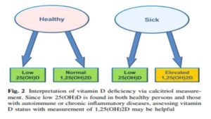
    What often presents, as “low vitamin-D” on a routine blood test is merely a sign of inflammation. It is worth noting that inflammation is a clinical sign of Mg deficiency.

    (Again, please see Deng et al 2013, linked above and also Weglicki & Phillips, 1992a).

    What is wildly confusing to patients, and their practitioners, is that taking more hormone-D supplements does not, and will not, correct this inflammatory process, as is illustrated in Figure 2 (below):

    Mangin, M., et al. (2014). “Inflammation and vitamin D: the infection connection”
    www.ncbi.nlm.nih.gov/pmc/articles/PMC4160567/

  3. Hormone-D Supplements are diabolic in their impact on our Immune System.
    1. Taking vitamin-D, which increases the synthesis of 1,25(OH)2D3 (active hormone-D), makes cells more sensitive to hydrogen peroxide (H2O2). It is worth noting that all inflammatory cytokines stimulate the increased production of H2O2, which is a known by-product of inflammation.

      Krane, S.M., et al. (1990). “1,25-Dihydroxyvitamin D3 increases the toxicity of hydrogen peroxide: the role of calcium and heat shock.”
      www.ncbi.nlm.nih.gov/m/pubmed/2226658/

    2. Taking vitamin-D dulls the immune system.

      Contrary to popular opinion, there is no “Causal Connection” between vitamin-D intake and improvement in either innate or adaptive immune function. An important finding from a recent study presents a clearer picture of this little discussed reality:

      Although, unlike Vitamin-D deficiency and rickets, a causal link between vitamin D deficiency and immune disorders has not yet been proven, there is compelling evidence in the laboratory of beneficial effects of 1,25(OH)2D3 in inflammatory diseases where Th1 cytokines have a pathological role.

      Please know that there is far more to this story than the simple-minded emphasis on ingesting more hormone-D!

      Wei R., Christakos, s. (2015). “Mechanism underlying the regulation of innate and adaptive immunity by vitamin D.”
      www.ncbi.nlm.nih.gov/pmc/articles/PMC4632412/

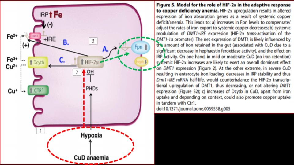
  4. Hormone-D Supplements are demonic by altering key genes involved in Iron metabolism.
    a) Contrary to popular belief in “high” vs. “low” thinking re nutritional metabolites, the body actually works off of optimal levels. Truth be known, our bodies run on a bell-shaped curve, which I prefer to call the “Goldilocks Syndrome.” (Far better to be at the peak, than at either pole!)


    Given that we’re now learning that vitamin D Is a regulator of Endothelial Nitric Oxide Synthase (eNOS), might the unchecked intake of this hormone be another important factor to consider in our metabolism getting out of balance and our genes getting *tweaked*

    Andrukhova O., et al. (2014). “Vitamin D Is a Regulator of Endothelial Nitric Oxide Synthase and Arterial Stiffness in Mice” 
    www.ncbi.nlm.nih.gov/pmc/articles/PMC5426652/

b) It’s worth noting that 20 years before learning about the impact of hormone-D supplements on eNOS, it was discovered that N.O. (Nitric Oxide) has a key regulatory role over IRPs (Iron Regulatory Proteins).

IRPs are an essential metabolite in directing iron metabolism function through their direct control of ferritin mRNA translation, as well as Transferrin Receptor-1 mRNA stability. This relationship of IRP<>IRE (Iron Responsive Elements) is among the most important, and sadly, among the most confusing aspects of iron metabolism.

Suffice it to say that in the absence of N.O., these IRPs act very differently, and that in the increased presence and prevalence of N.O., both the storage and absorption of iron, as well as the synthesis of hemoglobin are affected.

The net effect: Increased N.O. creates the illusion of iron deficiency by virtue of its impact to decrease the synthesis of ferritin, and increase the synthesis of transferrin receptors to absorb more iron.

That series of changes is a recipe for iron overload that presents as low ferritin, that wrong perception of low iron.

Pantopoulos, K. Hentze, M.W. (1995). “Nitric oxide signalling to iron-regulatory protein: Direct control of ferritin mRNA translation and transferrin receptor mRNA stability in transfected fibroblasts”
www.pnas.org/content/pnas/92/5/1267.full.pdf

Pantopoulos, K, Weiss, G., Hentze, M.W. (1994). “Nitric oxide and the post-transcriptional control of cellular iron traffic.”
www.sciencedirect.com/science/article/abs/pii/0962892494901791

c) And in a stunning study on this same metabolite, we learn that N.O. has an effect on 3 key genes — Post-Translationally — that are central to iron metabolism. These finding lend greater specificity to the findings of Pantopoulos & Hentze noted previously:

  • Decreased y-Globulin/eALAS function that affects Hgb synthesis;
  • Decreased H-Ferritin synthesis that affects storage of intracellular iron;
  • Increased TfR1 (Transferrin Receptor-1) that increases iron absorption

Domachowske, J.B. et al. (1996). “Nitric Oxide Alters the Expression of y-Globin, H-Ferritin, and Transferrin Receptor in Human K562 Cells at the Posttranscriptional Level”
www.bloodjournal.org/content/bloodjournal/88/8/2980.full.pdf?sso-checked=true

d) Finally, presented in this section are two key quotations from noted iron researchers, Guenther Weiss, PhD, William Hentze, PhD and colleagues, that serves to punctuate this profound physiological mechanism by N.O. that is little discussed, nor apparently properly understood in clinical circles.

A loose translation of this set of findings (#4a-#4d ^^^^) is that repeated supplementation with hormone-D appears to contribute to the oft declared diagnosis of “anemia,” that is overtaking the metabolic landscapes of patients worldwide.

It turns out that “low ferritin” and “low hemoglobin” may have a completely different origin than the habitually assumed declaration of low iron:

We conclude from these data that N.O. controls ferritin biosynthesis by changing the rate of translation of its mRNA

We have established that N.O. synthesized by macrophages and non-macrophages regulates IRF [today, known as “IRP”], the central Post-Translational Regulatory molecule of mRNAs involved in iron metabolism

Weiss, G., Hentze, W.M. et al. (1993). “Translational regulation via iron-responsive elements by the nitric oxide/NO-synthase pathway.”
www.ncbi.nlm.nih.gov/pmc/articles/PMC413641/

5. Hormone-D Supplements are destructive to Optimal Metabolism

a) In a study published over 55 years ago, it was revealed that vitamin-D supplements cause renal potassium wasting. Please know that depleting our cells of potassium, which is where potassium hangs out in our bodies, makes our cells increasingly vulnerable to an increase in intracellular iron that comes by way of high dietary iron and increased Transferrin Receptor activity as noted in the sections above.

Again, this is not a desirable mineral imbalance (i.e. low K vs. high Fe) in the short-term, and certainly not in the long-term!

Ferris T.F. et al. (1962). “Renal potassium–wasting induced by vitamin D” www.ncbi.nlm.nih.gov/pmc/articles/PMC291035/

b) Vitamin-D supplements cause ATP depletion and iron-mediated attack. Again, this appears to be a little-known effect of hormone-D supplements, but the impact to optimal metabolic function is stunning, especially in our kidney tissue.

Zager, R.A.,1999, “Calcitriol Directly Sensitizes Renal Tubular Cells to ATP-Depletion- and Iron-Mediated Attack”
https://www.ncbi.nlm.nih.gov/pmc/articles/PMC1866639/

c) Vitamin-D destroys vitamin-A reserves.
Mawson, A.R. (2012). “Role of Fat-Soluble Vitamins A and D in the Pathogenesis of Influenza: A New Perspective”
www.hindawi.com/journals/isrn/2013/246737/

key footnote [#96] in Mawson (2012) examines a never-discussed dynamic of vitamin-D levels affecting vitamin-A status and stores in the liver and the body. And despite what the title says on this study (Aburto et al., 1998), here is a key sentence from Mawson (2012) that is drawn from this critically important footnote:

“Although little is known about the effect of reduced sunlight exposure and/or deficient vitamin D levels on vitamin A metabolism, even small to moderate doses of vitamin D in chickens reduce liver vitamin A stores and lower the level of vitamin A in blood” [96].

I was stunned the first time that I read that sentence re the impact of vitamin-D on vitamin-A!

Does anyone honestly think that high intakes of this hormonal, supplemental-D would act any differently inside our (chicken-fed) bodies? Is anyone else just a little suspicious that vitamin-D is always described as being “deficient,” and vitamin-A is always found to be “toxic” in the literature?

Hmm?

Please know, this is the only study (Aburto et al, 1998) that even hints at this impact of hormone-D on vitamin-A. Know that I have been looking for validation of this fact for a number of years.

Aburto A, Edwards HM Jr., & Britton WM, (1998).  “The influence of vitamin a on the utilization and amelioration of toxicity of cholecalciferol, 25-hydroxycholecalciferol, and 1,25 dihydroxycholecalciferol in young broiler chickens,”

https://www.sciencedirect.com/science/article/pii/S0032579119408523

6. Hormone-D Supplements deflect the truth about what sunshine does for us.

a) Pine trees, grown in acidic (high iron) soil, produce copper rich products especially vitamin-C.
www.wildedible.com/pine-needle-tea-natural-vitamin-c

There are similar properties with citrus fruit trees, oak trees with acorns, pecan trees with pecans, etc. Wouldn’t it be valid to say that vitamin-C, too, is a valid candidate to be the “sunshine” vitamin, given the role that sunshine plays in this biological transmutation?

b) Sunlight activates POMC, which is a key catalyst to the PAM enzyme of the Hypothalamus that regulates 12 other key enzymes/hormones in our body. Again, it’s fascinating that this powerhouse of transactions is triggered by sunlight >> POMC:

Sunshine activates a gene called POMC, which in turn helps create melanin that determines skin color. This beneficial gene enhances sex drive (the endorphins or ‘happiness hormones’), as well as leptin, which helps burn fat and keep you thin.

Skin Biology, Pickart L. (2019) “Sun Health”
http://www.suntanscience.com/sunlightandhealth.php

c) Sunlight activates the catalytic breakdown of retinol that is key to its impact.

Given the worldwide obsession with storage-D status, it is both surprising and important to learn of the interdisciplinary and interactive nature of vitamins A<>D:

For instance, solar radiation has opposite effects on vitamins A and D, catabolizing vitamin A but increasing the concentration of vitamin D; the effects of the two vitamins are mutually inhibitory; retinoids regulate airway epithelial cell growth, differentiation, and gene expression…

And when you fully understand the myriad metabolic roles of vitamin A, and how it is the downstream metabolites from Retinol and Retinoids that have such profound effects on our metabolism, suddenly the role of sunlight in our lives (and livers!) takes on a much deeper and more important role than previously thought.

And what ubiquitous agent blocks these key retinol-related events? None other than “Sunscreen!” Please know, our skin “burns,” not from a lack of sunscreen, but a lack of fat-derived, Retinol.

Mawson, A.R. (2012). “Role of Fat-Soluble Vitamins A and D in the Pathogenesis of Influenza: A New Perspective” (Link listed above in 5c)

d) Then we learn that sunlight converts a key Cytochrome P-450 enzyme, eNOS, to enable the transformation of fat soluble vitamin-D <> vitamin D3 Sulfate, a water-soluble (i.e. usable) form of vitamin-D.

As I understand this process, it is only sunlight that can activate this critical enzyme mechanism. And the importance of this transformation is equally profound for our optimal health, as thoroughly explained in this series of interviews between Stephanie Seneff, PhD and Joseph Mercola, DO [No longer available]:

Regrettably, what is absent in this ^^^^ otherwise powerful and informative exchange between these two “greats,” is any consideration of the importance or the interaction with real vitamin-A, aka Retinol in this process. And why is this so important to our health

Because retinol, and its derivatives, are considered the “universal chromophore”, they have unique properties to harvest and harness light! There is a powerful connection between our eyes, and our liver, both organs being dependent on real vitamin-A, aka retinol.

Do we now have yet a third potential vitamin candidate to be the “sunshine” vitamin?

It is curious how the very start of the process to make vitamin-D depends on sunlight to activate a Cytochrome P-450 enzyme that requires copper, and that the critical VDR (Vitamin-D Receptor) is helpless without the direct involvement of a key Retinoic Acid Receptor, RXR derived from the metabolic breakdown of Retinol.

Musings of a mineral geek –

Zhong, M et al. (2012). “Retina, Retinol, Retinal and the Natural History of Vitamin A as a Light Sensor”
www.ncbi.nlm.nih.gov/pmc/articles/PMC3546623/#!po=0.515464

7. Hormone-D dulls our potential for Optimal Health

In what is regarded as the most robust Meta-Analysis of vitamin-D across a wide variety of conditions, a key summary comment from this analysis of some 187 other studies seems to identify the emerging and uncomfortable truth re the hormone-D hysteria gripping the planet:

Recent reports have highlighted the lack of concordance between observational studies and randomized controlled trials, concluding that vitamin D is more likely to be a correlate marker of overall health and not causally involved in disease.

When we were kids, the expression was “all bark, and no bite!”

Trust me, the dreary bloom is falling off this dietary daffodil!

Theodoratou E, Ionnidis JPA et al. (2014). “Vitamin D and multiple health outcomes: umbrella review of systematic reviews and meta-analyses of observational studies and randomised trials”
www.bmj.com/content/bmj/348/bmj.g2035.full.pdf

And just to solidify your understanding of the restrained “need” for vitamin-D, there is no clinical benefit to having storage-D (25(OH)D) >21ng/dL. This was the sterling and unsettling conclusion of a recent study to contrast vitamin-D status against all-cause mortality.

This figure flies in the face of the absurd Lab ranges for storage-D that start at 30 and go up to 100! The 2014 Study noted ^^^^ above is a chilling indication of how out of touch the meme is about hormone-D, which is amplified in Dr. Amer’s equally deflating conclusions below.

Amer, M. et al. (2013). “Relationship between 25-Hydroxyvitamin D and All-cause and Cardiovascular Disease Mortality”
www.amjmed.com/article/S0002-9343(13)00084-3/pdf

Ok, that is a lot of info. Please forgive the download.

I realize that this post is a doozy, but I’ve been collecting these articles for months, and decided it was time to do the definitive Vitamin-Dump!

Please know, I will be drawing upon several of these studies for posts in the next several weeks/months as I plan to do a series of Posts to drill deeper into the 2nd and 3rd order implications of these dynamics.

So, let’s drop the pretense. There is a great deal more to this synthetic, soy-based (not sun-based) toxic supplement than you are being led to believe on the Internet and in your doctor’s office.

It is not a panacea supplement for improving health.

In fact, following my close to 10 years of analysing and evaluating this issue, I would argue that the use of hormone-D supplements represents a central pathway to weaken our mineral dynamics, our metabolic pathways, and ultimately, our immune system.

And finally, an important caution, please beware of studies that are based on Correlation, and not Causation. A critical question to ask yourself, and to amplify this point: “Do flies cause garbage?”

Of course not! And this is true of hormone-D, too!

It appears that “Low vitamin-D” shows up (i.e. it is correlated) with countless conditions, but its lack is hardly cause of these many conditions, and its supplementation is hardly a correction of these same conditions. (Please take a moment to re-read that sentence, once again) Worse yet, it seems that the much bally-hooed “benefits” of taking this synthetic supplement are more illusory, than real. Why are we not surprised?

In conclusion, supplementing with this “poison,” because you think you have a dearth of this storage hormone in your blood, could very well be the stimulus for the death of your vitality, in ways you never imagined possible.

“A” votre sante!
Morley M. Robbins