Iron Toxicity Post #62: You do NOT have a Candida Infection, you DO have an Iron Infestation!

Iron Toxicity Post #62: You do NOT have a Candida Infection, you DO have an Iron Infestation!

You do not have a Candida Infection. You do have an Iron Infestation!

Today’s post will be short and sweet. I think you will be stunned at the clarity of the iron-based research, re. Candida albicans, as well as the brevity of my message about addressing the iron root of this problem!

In my never-ending quest to bring you the truth about the role that excess, unbound iron plays in causing all known conditions, disorders, syndromes, and dis-eases. You will absolutely love what one of my magnesium heroes, Ronald J. Elin, MD, PhD (University of Kentucky Medical Center), has to say about the iron origin of candida albicans:

Elin R.J., Wolff S.M. (1974). “The Role of Iron in Nonspecific Resistance to Infection Induced by Endotoxin.”
pdfs.semanticscholar.org/de95/2a2d800e6e233a7fae838b260e2d15210bea.pdf

There are some important quotations from this penetrating study, the role that iron plays in supporting and enabling a yeast infection:

“Since it is known that Iron is an essential growth factor for C. albicans [14, 15] and that endotoxin [e.g. LPS, aka Lipopolysaccharides from Bacteria] causes [emphasis added] a significant fall in serum iron [16], the importance of iron as a factor in nonspecific resistance to infection with C. albicans induced by endotoxin was evaluated in vitro and in vivo.”

Know that infections and inflammation causes hypoferremia. The innate immune system is designed to sequester iron away from the critters.

“The step-like pattern of this dose-response experiment clearly demonstrates the in vivo importance of Iron in C. albicans infections.”

As I have been saying for close to three full years, all pathogens (whether they be, bacteria, fungus, virus and parasites) exist, replicate and thrive on an iron buffet. It is the only thing that Louis Pasteur, PhD forgot to tell us, as he chose to terrorize us with fostering an absurd fear of bugs, critters, and chiggers, etc. and not the metal that they are attracted to. This research and the figure below seal the deal. It’s a compelling piece of causal research!

I wish I had a dollar for every client in the past 9yrs:

  • Who had a yeast infection
  • Who was totally stressed out about it
  • Who was clueless about iron’s role in its formation
  • Who was taking toxic Rx meds to attack it and
  • Who was doing nothing to address the underlying causative iron overload supporting the very inception and lifespan of this so-called yeast infection?

What to do?

Please, stop attacking the guest! Taking fluoride-activated anti-fungals” are not the answer!

Please start strengthening the host!
https://therootcauseprotocol.com/about/
Please consider getting this full Monty Iron panel:
https://therootcauseprotocol.com/order-lab-tests/

Please know that the body has amazing powers of resilience when it is properly nourished! I truly hope that those dealing with this latest clinical sign of iron overload can see and act on this truth.
 
A votre sante!
Morley  M Robbins

For Facebook Discussion:
https://www.facebook.com/groups/MagnesiumAdvocacy/permalink/1448954445172620/

Iron Toxicity Post #61: EYE-Ironic origin of Alzheimer’s disease

Iron Toxicity Post #61: EYE-Ironic origin of Alzheimer’s disease

This post is about Eye/Iron or Eye-ronic origin of Alzheimer ’s disease and all other forms of neurodegeneration known on this Planet.

“Discovery consists in seeing what everybody else has seen and thinking what nobody has thought.”— Albert Szent-Gyorgyi, 1937 Nobel Laureate: “Discovery of Vitamin ‘C’ complex”

I have wanted to write this post since the beginning of 2017, but several key pieces of the puzzle were cleverly eluding me. Please know that I am far from done understanding all of these dynamics of neurodegeneration, but I am reasonably confident now to make several key assertions, which I’ll do throughout this post with a wrap-up at the end.

I am quite confident; many of you will not entirely like what this post has to say. You will be challenged to understand the technical aspects of it and I barely have a full grasp. But the mineral and metabolic issues that are covered in this post will grow in importance, and will become a key cornerstone in our understanding of how best to get out of our iron shackles and regain our metabolic homeostasis.

Some notable facts about neurological conditions, in general, and neurodegenerative issues, in particular:

  • One billion people, worldwide, nearly 1 in 6, suffer from neurological disorders: AD, PD, MS, strokes, epilepsy, migraines, brain injuries and other forms of neuroinfection.
  • Thirty seven million people worldwide suffer with Alzheimer’s
  • Ten million people worldwide suffer with Parkinson’s

We all know at least 1-2 people (family members, friends, work associates, neighbors, etc.) that are being ravaged by the information that surrounds these brain-related neurodegenerative conditions. It is absolutely time for this level of clinical inadequacy to stop. 

I’m doing my part today by sharing this extensive panel of research to compile the leading thinking in a way that presents what I regard as a compelling argument about a major destructive factor that is right under our noses. Which is contributing to the unchecked acceleration of neurodegeneration and misery on this planet.

So what’s really going on here with these neurodegenerative diseases?  Isn’t that what you’ve come to expect to learn innovative and foundational metabolic nuggets from these “Insightful Teaching Posts” (otherwise known as Iron Toxicity Posts)?

  • It’s important to know that Alzheimer’s disease (AD) is characterized by the following mineral patterns in the brain tissue:
    – High copper,
    – Low iron,
    – Low zinc, typically
    – Low ferroxidase function (ceruloplasmin),
    – High oxidized serotonin, especially in the hippocampus
    – Confusing reference(s) to Monoamine Oxidase (MAO, as a bad guy) that is meant to “turn off” neurotransmitters
    – Silence on melanin’s role in the locus coeruleus that affects the hippocampus
  • It’s equally important to know that Parkinson’s disease (PD), on the other hand, is characterized by these mineral patterns in the brain tissue:
    – Low Copper,
    – High iron,
    – Low zinc, typically…
    – Low ferroxidase function, once again,
    – High oxidized dopamine, especially in the substantia nigra
    – Confusing reference(s) to MAO (as a “bad guy”), again, that is “turning off” the neurotransmitters
    – More silence on melanin’s role in the locus coeruleus that affects the substantia nigra.

Clearly, there are some notable and contrasting mineral dynamics, which adds to the mystery and clinical confusion about these two most prevalent forms of neurodegeneration on the planet. 

But please understand the fact that both conditions, and all neurodegenerative conditions, collectively, that I’ve studied, have a notable lack of ferroxidase function.

This is a critical point to which we will return shortly.

Okay, so with that as a background, let’s begin this blinding blitzkrieg blog!

I’ve known for quite some time that EMFs, you know those pesky little electromagnetic frequencies that every conceivable device we use emits has an effect on our iron status. In effect, this is a ubiquitous form of environmental stress. What we’ve learned is that there is always a Magnesium Burn Rate (MBR) attached to any form of stress. 

EMFs are no different in their Magnesium (Mg) depleting effects, and if Mg goes down, trust me, the toxicity of iron is going to be going up! Here are several blockbuster articles for those of you seeking to better understand this EMF dynamic to get a better handle on how this tidal wave of radio frequencies is altering not only our external environment, but also our internal antioxidant enzyme landscape:

Maaroufi, K., et al. (2014). “Spatial learning, monoamines and oxidative stress in rats exposed to 900 MHz electromagnetic field in combination with iron overload”
pdfs.semanticscholar.org/29a3/7abf4734218e0e6276afd52ff04c59650562.pdf?_ga=2.68949969.1975053235.1498943746-1468208896.1498943746

Gherardini, L., et al. (2013). “Searching for the Perfect Wave: The Effect of Radiofrequency Electromagnetic Fields on Cells” (The impact on Complex IV, cytochrome c oxidase is particularly noteworthy)
www.ncbi.nlm.nih.gov/pmc/articles/PMC4013569/

Consales, C., et al. (2012). “Electromagnetic Fields, Oxidative Stress, and Neurodegeneration”
www.hindawi.com/journals/ijcb/2012/683897/citations/

Please note that the first study noted (Maaroufi, 2014) is a very provocative one that chose to combine EMFs (900 MHz) with iron loading on the experimental rodents. The sentence that really caught my “eye,” (pardon the pun) was the following:

“A link between EMF, iron accumulation in the brain and neurodegenerative disorders including Parkinson’s and Alzheimer’s diseases has been suggested.”

Are you sitting up a bit straighter in your chair right about now? So, I’ve known about these iron properties of EMFs for quite a while, and I knew that there had to be a connection to the use of blue light that many of these devices emit, but I lacked the causal connection between that high-energy spectrum of light and its impact on iron. There is an absolute relationship between copper and the copper-dependent enzymes [think cytochrome c oxidase] and the other low energy end of the light spectrum where red and infrared light hangs out.

Everything changed a couple of weeks ago when my friend and colleague Alicia Barone Stephens happened to post an article about “Blue Light” and “Alzheimer’s” that had some critical commentary & insights from brain surgeon, Jack Kruse, MD. 

“Culprit in Plain sight in Alzheimer’s disease development”
medicalxpress.com/news/2017-06-culprit-hidden-plain-sight-alzheimer.html

Jack Kruse, MD commenting on Facebook:
www.facebook.com/drjackkruse/posts/1798230000241346

And I quote from Jack Kruse, MD:

“Too bad no one in this paper realize that transition metals, like Iron, draw nnEMF [non-native EMF] toward them because of the D shell electrons they contain. That is why AD is blowing up in a blue-lit microwaved world. CULPRIT HIDDEN IN PLAIN SIGHT IN ALZHEIMER DISEASE DEVELOPMENT”

It doesn’t get too much clearer than that. I’m delighted that Jack Kruse, MD elected to connect these dots so precisely. What is vital to understand is that our eyes are rich sources of iron to support the high metabolic demands of our eyes, and yet this pesky heavy metal accumulates as we age. This is not my conjecture; it is addressed in almost every article that I’ve read about eye disorders and disease over the last two years.

Here’s one of ~35 that I’ve read on this eye/iron dynamic:

Loh A. et al. (2009). “Iron homeostasis and eye disease”
www.ncbi.nlm.nih.gov/pmc/articles/PMC2718721/pdf/nihms-125393.pdf

Okay, so what do we know thus far?

  • The incidence of neurodegeneration is exploding all over this planet.
  • We know that the two most prevalent forms, AD and PD, have notable mineral differences, yet notable overlaps in key enzyme dynamics that cause these so-called conditions.
  • We know that our contemporary and constantly connected environment is teeming with technology that is causing both oxidative stress, and has known connections to the very heavy metal that fuels the relentless oxidative stress that ails us.
  • And while we’ve talked about this iron-induced oxidative stress many times before, the spin has always been around our food and dietary intake of enriched iron not our eyes!

What I want to emphasize in this iron toxicity post #61, is that there are added nuances to this crisis that even I was unaware of until forcing myself to sit down and piece this puzzle together. And I’m hardly done, despite all the hours, and hours of effort to read, synthesize and now write this unsettling piece. 

As it turns out, our daily intake of our popular and dependent technology is only adding to our growing metabolic woes, especially as we age. Trust me, I would much rather be outside enjoying this beautiful Saturday (July 1, 2017) than be zapping my eyes, my thyroid (thank you Jack Kruse for that keen nuance that I missed!), as well as my brain, with this nnEMF/Blue Light emitting, oxidative stress-creating computer screen!  But I figure you all are worth it!

Alright, if there is anyone at this point that doubts that the foundational flaw in the conventional understanding of the meteoric rise in the prevalence of AD or PD has nothing to do with a lack of ferroxidase enzyme function to keep oxidative stress in the brain at bay, please note the following studies:

A sampling of proof that Alzheimer’s and Parkinson’s are connected to copper<>iron dysregulation:

Kristinsson, J., et al. (2012). “Ceruloplasmin and iron in Alzheimer’s disease and Parkinson’s disease: a synopsis of recent studies”
www.ncbi.nlm.nih.gov/pmc/articles/PMC3493298/

Torsdottir, G. et al. (2011). “Ceruloplasmin and Iron Proteins in the Serum of Patients with Alzheimer’s Disease”
www.ncbi.nlm.nih.gov/pmc/articles/PMC3243634/

Montes, S., et al. (2014). “Copper and Copper Proteins in Parkinson’s Disease”
www.hindawi.com/journals/omcl/2014/147251/

These are merely three of ~50+ articles that I have read and analysed to get to the root cause of this maddening and frightening neuro-crisis. Up to this point, I was quite clear and confident that the liver was the single greatest producer of ferroxidase enzyme (FOX, or ceruloplasmin oxidase, or Cp), and that the brain was #2. That hierarchy had been noted in dozens of recognized and repeatedly referenced articles. It is worth noting that FOX is also made in the endocrine glands, the kidney, the uterus, the genitals, the breasts, etc., anywhere there is high metabolic activity to address the potential toxicity of iron found in oxygen-rich haemoglobin.

Everything changed when I read this mind-numbing study by Lin Chen, and colleagues at the University of Pennsylvania, at the start of this year:

Chen, L., et al. (2003). “Increased Expression of Ceruloplasmin in the Retina following Photic Injury”
Abstract: iovs.arvojournals.org/article.aspx?articleid=2416930
Full Article: www.molvis.org/molvis/v9/a22/v9a22-chen.pdf

Please take a moment to familiarize yourself with these findings and let the significance of this study sink in, in light of all that we’ve covered, especially these past two years. What Dr. Chen and colleagues discovered is that the eyes make 8 x more ferroxidase enzyme (ceruloplasmin) than the brain. Do you understand the magnitude of these findings? That it is our eyes that are not only taking it “on the chin” with the increased oxidative stress, but that they are supposed to be making eight times more ferroxidase than our brains in a nutritional/medical environment that has gone out of its way in the past 50+ years to destroy the body’s and the eye’s ability to make ferroxidase enzyme. Then on top of that, add insult to injury by going on to not even bother to measure it. (Thus, the infamous “not known, and not looked for” medical mantra)

Do you finally understand why I despise hormone-D so much? That obnoxious, synthetic, toxic supplemental hormone that is completely misunderstood by scientists, clinicians and the masses kills retinol, the very metabolic backbone for making this vital ferroxidase enzyme.

Isn’t it interesting that we are chided for using retinol, even though it is essential for the very function and metabolic activity of the retina? Get it? Retinol for the retina! Do you now finally understand my obsession with the Root Cause Protocol, is inspired by Mother Nature, is designed to enhance the viability and function of the ferroxidase enzyme (FOX, ceruloplasmin oxidase, and Cp)?

In case you think that that retina<>ceruloplasmin was a “one-and-done-study,” here’s what that provocative team based at UPenn went onto discover, in addition to their blockbuster revelations about FOX:

Hahn, P., et al. (2004). “Disruption of Ceruloplasmin and Hephaestin in mice causes retinal iron overload and retinal degeneration with features of age-related macular degeneration”
www.ncbi.nlm.nih.gov/pmc/articles/PMC518844/pdf/10113850.pdf

So, here we have proof that the very disruption of ferroxidase function, as expressed in both ceruloplasmin and hephaestin (an enzyme cousin of Cp), causes iron to build, which then goes on to create even more oxidative stress.

So, has anyone been wondering why I chose the graphics for this particular Post?

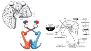
What we are seeing in those pictures is the unique structure and placement of the eye, it’s optical nerve and the amazing optic chiasm [Greek for “criss-cross”] that creates an “X” in a particularly important and vulnerable part of the brain:

  • The pituitary & thyroid are below that chiasm.
  • The limbic region of the brain (think master regulatory regions, like the hypothalamus, etc.) is directly above that chiasm.

Do you think that there just might be building, wandering, and destructive iron-induced oxidative stress that is associated with the retina(s), and subsequently the optic nerve(s), that is compromised in its ability to make the very antioxidant enzyme that must be vitally important for our optimal health? Or why would our maker have enabled us to make so much of this antioxidant enzyme, and in that particularly sensitive area of our body?

It would be reasonable to think that the systemic and progressive lack of this ferroxidase thingy is the sole reason for all this chaos, but as Mark Twain might say: “It just AIN’T so!” There are other antioxidants involved, as well. There are brain chemicals known as prion proteins, and it was just today that I learned that they are copper dependent. I had not learned that in my prior readings. These key proteins play very important roles as an antioxidant, but they get functionally tweaked, especially when they’re in the presence of iron-laden macrophages, that are called microglia and astroglia, which are found in the brain region.

Here are some compelling studies that support the role of prion proteins, but also how they are being accused, wrongly, in my humble opinion, for being a source of the problem, when they are seeking to come to the aid of the oxidizing tissue. Nothing like blaming a key component that is being affected by iron-toxic macrophages that alter the optimal function and physiology of these copper-driven chemicals.

Price, D.L., et al. (1993). “Alzheimer’s Amyloid Plaque and Prion Diseases”
www.ncbi.nlm.nih.gov/pmc/articles/PMC46935/pdf/pnas01471-0012.pdf

Kercher, L., et al. (2007). “Prion Protein Expression Differences in Microglia and Astroglia [Macrophages] Influence Scrapie-Induced Neurodegeneration in the Retina and Brain of Transgenic Mice”
jvi.asm.org/content/81/19/10340.full.pdf+html

Zhang, W., et al. (2014). “Role and mechanism of microglial activation in iron-induced selective and progressive dopaminergic neurodegeneration”
www.ncbi.nlm.nih.gov/pubmed/24277523

It does not stop there, unfortunately. Anyone who has loved ones suffering from these neurological conditions, especially Alzheimer’s, is aware of amyloid protein precursor (APP) that, as it turns out, actually has ferroxidase enzyme function. APP is seeking to quell the rising oxidative stress that is coming from the daily intake of iron-enriched food and our never far-from-us iron enriching electronics!

Duce, J.A., et al. (2010). “An iron-export ferroxidase activity of β-amyloid protein precursor is inhibited by Zinc in Alzheimer’s Disease”
www.sciencedirect.com/science/article/pii/S0092867410009384

www.ncbi.nlm.nih.gov/pmc/articles/PMC2943017/

Take stock of the added significance of this study by noting that zinc inhibits the ferroxidase activity of this critical brain tissue antioxidant. This is consistent with other studies that have proven that zinc zaps ferroxidase function, which we have examined in prior posts. Please, do not use zinc supplements, under any circumstances, if your ultimate objective is to increase the level and functionality of your ferroxidase enzyme.  You can get zinc from your diet, not from a bottle.

Let’s keep it simple, seekers.  Let me quote the last sentence of the abstract of this illuminating study by Duce, 2010:

“Iron causes selective and progressive dopaminergic neurodegeneration, and microglial NOX2 activation potentiates the neurotoxicity. PKC-σ, P38, ERK1/2, JNK, and NF-КBP65 are the potential molecules relevant to microglial NOX2 activation.”

It would be wonderful if more practitioners, pundits and FB posters would be this direct about all this clinical chaos occurring inside the brains of their patients and readers!

The three most dangerous minerals, in a supplemental form, that are the mainstays of allopathic practitioners are calcium, iron and zinc. They are not the answer, they have never been and they never will be. I only hope that more readers will have fresh eyes to see the mineral truths in this post #61.

Foundational iron hypothesis:

“Iron and iron dysregulation, due to a systemic lack of bioavailable copper, causes neurodegeneration period!”

What is rarely, if ever addressed, are the many key brain and neural tissue antioxidants that are being under produced and/or compromised time, and time again, throughout the brainstem and the brain cortex tissue. 

We come to learn that it starts in our eyes! The bulk of conventional neurodegeneration research blows entirely past the central role of ferroxidase enzyme, prion proteins as key antioxidants, APP as yet another key antioxidant enzyme. What I am now discovering is that there is a granddaddy anti-oxidant of them all, it’s called melanin.

Melanin (pigmentation) is found in hair and skin and guess what, it is found in the eyes too.  It is not about the colour of the eye and no, I am not talking about melatonin, I am zeroing on melanin.  Melanin has a key chemical that plays a central role in many areas of the brain, not the least of which is the locus coeruleus [which means black dot], which is a major source of making dopamine to influence the functionality and balance of many, many sections of the brain.

Arias-Esparza, M., et al. (2011). “The Unexpected Capability of Melanin to Split the Water Molecule and the Alzheimer’s Disease”
https://www.scirp.org/journal/PaperInformation.aspx?PaperID=7404

This study above study is an absolute must read. This study breaks critical new ground, and shatters the very foundation of our known knowledge of cellular energy, electrical transmission around the body, as well as key anti-rusting factors, to name but a few functions, that have been shielded from us. I know not why, but I have a few theories.

No, melanin is not just skin, and hair pigment! 

But for those that want to fully delve deeper into this, I would strongly encourage you to read the following:

King, R., M.D. (2001). “Melanin: A Key to Freedom”
www.amazon.com/Melanin-Key-Freedom-Richard-King/dp/1602810958/ref=sr_1_12?s=books&ie=UTF8&qid=1498928462&sr=1-12&keywords=melanin

Bynum, Bruce, E. (2014). “WHY DARKNESS MATTERS: (New and Improved): The Power of Melanin in the Brain”
https://www.amazon.com/WHY-DARKNESS-MATTERS-Improved-Melanin/dp/1502411172/ref=sr_1_5?s=books&ie=UTF8&qid=1498928462&sr=1-5&keywords=melanin

I plan to write much more about melanin in future posts, but suffice it to say we have been completely misinformed about:

  • Its role as a source of mitochondrial energy,
  • Its role as a super conductor of electricity – it is prevalent in key areas of the central nervous system (CNS).
  • Its role as an iron-trapping agent, which is why its prevalence builds in lock step with the rising iron in the eyes.
  • Its role as a master absorber of the complete visible light spectrum.
  • Its role as a master absorber of sound in the inner ear.
  • Its role as a critical facilitator of pineal health and function.
  • Trust me, I could go on and on.

What is particularly poignant to understand is that melanin, in my humble opinion, has been hijacked and relegated to this lowly status as a pigment. It has also been methodically corralled into a master iron-trapping agent, which has completely diverted its ability to fulfil its majestic roles in the human body, as briefly and incompletely noted above.

In effect, melanin has become the “iron police force” responding to the tidal wave increase of iron in our bodies. Melanin is most effective at locking up that toxic heavy metal iron, and keeping it out of harm’s way. Here’s a clever twist, what’s a popular nickname for Policemen?  Yes they’re called coppers! So we’ve got coppers putting this toxic metal behind bars! 

How do you make melanin? The synthesis of melanin is entirely dependent on another key copper enzyme: Tyrosinase!

Where is that enzyme found in Mother Nature?

Why none other than wholefood vitamin-C that Albert Szent-Gyorgyi, PhD discovered in the 1930’s studying peppers from his hometown of Budapest, Hungary!

The most effective way to bind up melanin is with absurd levels of iron fortification in the food.

The most effective way to prevent melanin production is to drown the masses with ascorbic acid (devoid of any tyrosinase enzyme) and synthetic hormone-D that drives iron storage deeper and deeper into the tissue, which also causes renal potassium wasting (Ferris, 1962) thereby making the cells and the tissue more attractive to iron storage, and iron-induced oxidative stress!

Ferris, T.F., et al. (1962). “Renal potassium-wasting induced by vitamin D”
https://www.ncbi.nlm.nih.gov/pmc/articles/PMC291035/

The most effective way to distract or detract melanin entirely from the scene, and guarantee global neurodegeneration, is to do just what the system has done. As Jack Kruse, MD so aptly states, it’s been done right under our noses and we never even saw it!

Well, I’ll just speak for myself, I never saw this full court press of multiple iron factors to ensure neurodegeneration coming, despite my years of meticulous, minerals-based research. We can no longer ignore these conclusions.

So we learnt the saying is apt:

“Not known, because not looked for”

Okay, so what to do?

  • Stop believing anything anyone has to say about neurodegeneration being a medical disease. (This is complete and utter fantasy on their part, and clearly a lack of awareness of the literature)
  • Start to focus on understanding that excess, unbound iron, and the dysregulation that iron creates, is the very epicenter of this destructive metabolic dynamic.
  • Stop thinking or believing that this is a genetic condition or a familial condition, that is simply not so, it is entirely an epigenetic one, or said another way, an environmental condition.
  • Start understanding the scope and reach of iron-induced oxidative stress in all aspects of conditions and syndromes that you might be aware of or battling. They are truly all connected and riding on that “Ferrous Wheel” that runs your symptoms.
  • Stop, believing that you are anemic! (Nothing could be farther from the truth)
  • Please start the Root Cause Protocol to reverse this relentless pattern of mineral dysregulation that is triggered by iron.

I do apologize for the length of this particular post. It has been building for 6+ months, and I have wanted to share these important insights and discoveries for that entire time, but I also wanted to wait for just the right moment, when I felt more complete, and thus more confident, in my observations and conclusions.

This is our time! We absolutely need to pursue our health freedom, if for no other reason than to gain our medical independence from misguided clinical thinking that still believes neurodegeneration is a medical disease despite the countless articles that I’ve cited!

I trust that those of you who have labored and hung with me throughout this entire post would agree that that kind of clinical belief is seriously flawed, at best, and grossly irresponsible, at worst.

In subsequent posts and on-line chats, we will explore the important steps and options that we can all take to curb this iron influx of toxicity in our gut, and now as we’ve learned, our eyes.

A votre sante!
Morley M Robbins

For Facebook Discussion:
https://www.facebook.com/groups/MagnesiumAdvocacy/permalink/1400311870036878/

Iron Toxicity Post #60: ‘Am I really anemic?’ or said another way: ‘Not known because not looked for’

Iron Toxicity Post #60: ‘Am I really anemic?’ or said another way: ‘Not known because not looked for’

“Am I really anemic” Playbook or said another way: “Not known because not looked for”

When last we met during Iron Toxicity Post #59:
https://www.facebook.com/groups/MagnesiumAdvocacy/permalink/1366914676709931/ 
I developed what I believe (& hope that you agree with) is a compelling set of facts to seriously question the validity and the clinical relevance of ferritin-only blood tests as a stand-alone indicator of true metabolic iron status in the human body.

In that post, I noted that there were two more posts coming, one on key questions to ask, and an additional post on suggested tests to broaden our understanding and assessment of the other factors involved in iron metabolism. In the spirit of keep it simple seeker (K.I.S.S.), I have elected to simplify this process. I am going to blend the two pending posts, which is the focus of this set of comments and insights in this current post #60.

Before proceeding, let me amplify the second half of the title: “Not known because not looked for.”

We are led to believe that the medical system is exhaustive in its blood-testing search for clues to what’s not right with our metabolism. What I have come to discover is that much of the laboratory testing, that is commercially available around the world, is based far more on availability, not so much on clinical accuracy or applicability.

An obvious case in point: No one tests for Magnesium RBC, despite its obvious importance in all 100 trillion cells, as well as some 3, 751 proteins known as the magnesome.

Another more obvious point: No one tests for ferroxidase enzyme function, the master anti-oxidant enzyme in the human body that no one seems to know about.

These are but two key tests, among scores of “missing” tests from the typical arsenal that doctors are trained to provide, and allowed to order. Though this group of available tests does little to meaningfully pull back the curtain on the true mineral dysregulation that is driving the cellular dynamics of metabolic dysfunction.

At the end of the day, all we can do is the best that’s currently available, but that should not deter us from pointing out the glaring shortages and inadequacies of current laboratory testing for iron status, that drive inconclusive, and at times, faulty clinical conclusions.

What now follows is my best recommendations for a 10-point plan for “Questions to ask,” and where appropriate, “Tests to demand,” when confronted with the oft-repeated phrase: “You’re anemic!” I strongly advise you all to stand your ground and get the truth of what’s really driving your iron markers. I trust that you will find the following information, both useful and life-changing:

Q. 1) Doctor, given that ferritin is not synthesized in the blood, exactly how does a serum measurement of ferritin (i.e. an extracellular marker, and akin to “kitchen temperature”) relate to, or have any relevance to, the intracellular ferritin protein level (i.e. the storage protein synthesized Inside the cells, and is more akin to “oven temperature”)?

Recommended test: There is no known measure of intracellular ferritin.

Q. 2) Given that there are two forms of ferritin, what form(s) of ferritin are present in this serum measurement: ferritin-h (heavy, with ferroxidase enzyme function) and/or ferritin-l (light, without the key ferroxidase enzyme function)?

Recommended test: Distinction of ferritin-h or ferritin-l (this is a standard test in research studies, but I’m not certain that this test is even available commercially. the intent here is to drive home the point that a ferritin-only test is spurious, and misleading, at best).

Q. 3) Doctor, what’s the status of my hemoglobin, given that ~80% of my body’s iron is complexed in that key oxygen transport protein — as opposed to ferritin that has ~10% of the body’s iron?
Recommended test: Hemoglobin
requestatest.com/hemoglobin-testing

Q. 4) What is the status of my mineral and vitamin co-factors that regulate iron metabolism: i.e. magnesium, bioavailable copper, zinc, ceruloplasmin (ferroxidase enzyme), and several select iron markers?

Recommended test:
requestatest.com/mag-zinc-copper-panel-with-iron-panel-testing
https://requestatest.com/mag-zinc-copper-panel-with-iron-panel-plus-vitamin-a-and-vitamin-d-test

Q. 5) I realize that all you’re seeing in this “low ferritin” is “anemia of iron deficiency” (AID), but is there a chance that I could, in fact, be dealing with “anemia of chronic inflammation” (ACI)? (It is a well-established fact that inflammation causes “hypoferremia,” i.e. low iron in the blood)

Recommended test: Markers of inflammation: bun/creatinine ratio; hs-CRP; IL-6, blood glucose; Hgb A1-c; etc.

Q. 6) Exactly how would you know about this distinction between AID and ACI if you haven’t done these additional tests that assess inflammation? In an inflammatory process, wouldn’t we expect to see an elevated IL-6, but that if there were a true iron deficiency, IL-6 would not be elevated? Wouldn’t that be a decisive indicator of true iron status?

Recommended test: Interleukin-6; Interleukin-1; TNF-a; and NF-kB (These markers are routinely assessed in research studies, but I’m not sure about their availability with the commercial labs)

Q. 7) Is it possible that my low level of bioavailable copper, as expressed by low ceruloplasmin (Cp, as known as ferroxidase), or suspiciously high Cp, could be causing “iron retention” in the macrophages, thus impairing proper iron egress from the cells in my body?

Recommended test: Stress the importance of the serum ceruloplasmin test as a sign of bioavailable copper.

Q. 8) Doctor, is it possible that my hepcidin level, the iron peptide hormone, could be elevated which would then prevent iron from being released from:

  • Enterocytes in the digestive tract?
  • Hepatocytes in the liver (key to blocking iron recycling)
  • Macrophages in the iron R.E.cycling system, how would we know if we don’t test the status of this key iron hormone?

Recommended test: This is routinely done in research studies, but it is not available, to my knowledge, in a commercial lab.

Q. 9) Given that hepcidin is a “Janus (2-faced) peptide”, serving as both an iron regulatory, as well as anti-microbial agent, wouldn’t it be critical to our understanding hepcidin status before assuming “iron deficiency,” and especially before “iron supplementation,” given that:

  • Hepcidin increases with iron supplementation, as well as with the conditions of both infections and/or inflammation (Note: increased hepcidin expression causes “hypoferremia”)
  • Hepcidin expression decreases with increased need for iron in the body. Therefore, we expect low hepcidin expression if there were a true need of iron.

Recommended test: This is routinely done in research studies, but it is not available, to my knowledge, in a commercial lab.

Q. 10) Doctor, given my long-standing litany of symptoms and issues, how many of them are, for a fact, caused by iron-induced oxidative stress? I’m curious if you’ve had a chance to read any of the 59 Posts on Iron Toxicity that I shared with you recently?

Recommended link:
https://therootcauseprotocol.com/category/research/iron-toxicity/

I think you all get my point that assessing true iron status is a far cry from measuring your height or your weight. It is a Rubik’s Cube of interlocking factors that do not lend themselves to “one and done” testing.

Furthermore, as you can see, even the most sophisticated commercial labs are a veritable “swiss cheese” of blood tests, many of which are not available. As noted at the outset: “Not known because not looked for” That does not mean that if they are not available, that they are not relevant to your situation, especially given the importance of knowing your true iron status. In fact, I would contend that it is just the opposite.

This testing “patch-work” is a classic case of designed & selective awareness. Given that most of these tests are routinely done in research studies, around the globe, underscores their clinical importance, as well as their significance. In my humble opinion, it raises countless red flags why they are not available to your practicing physician(s) and to us as a group of individuals seeking to know the truth. I’ve got some theories on why this is the case, but I’ll let you draw your own conclusions.

I am also aware of the inherent “discomfort” that this series of questions raises for each of you in terms of your ongoing relationship with your healthcare practitioner. 

My intention is to put a spotlight on the set of variables and clinical factors that are essential for properly assessing and concluding that you are dealing with “iron deficiency,” and not “chronic inflammation!” And if your practitioner is resistant to your well-informed questions, you may want to consider seeking out a more open-minded healthcare provider. 

This raises serious questions about what they know, and what they want you to know. So, I do hope you find this set of questions helpful, and that it reinforces the points that I’ve made in my many, many posts on Iron Toxicity

A votre sante!

Morley M Robbins

For Facebook Discussion:
https://www.facebook.com/groups/MagnesiumAdvocacy/permalink/1385659851502080/

Iron Toxicity Post #59: A Tale of two irons and the hepcidin peptide that regulates them

Iron Toxicity Post #59: A Tale of two irons and the hepcidin peptide that regulates them

This post is based on a tale of two irons, hepcidin peptide that regulates them and causes chaos, confusion, clinical conditions and clinical currency.

“People would rather believe a simple LIE, than the complex TRUTH…”
— Lao Tzu (~600 BC)

For all intents and purposes, this post on iron toxicity is a Modern-day Survival Guide in an era when the only clinical and robotic response to “low iron blood markers” is ignorance, apathy and more iron. 

This post is = complex truth.

Healthcare providers wanting to give you more iron based on a ferritin-only blood test = simple lie

Famous Chinese curse: 

“May you live in ‘interesting times“ 

Well Dorothy, we’re not in Kansas anymore, but we are living in most interesting times.

This post will be among my last to put to bed this absurd and entirely engineered crisis about iron, copper<>iron metabolism. 

These last 3 posts will be in three parts:

  • Part I: The facts of iron anemia and what it really stands for: “low functional iron!”
  • Part II: Several key and salient questions that you need to ask before taking more iron!
  • Part III: The specific blood tests that must be done before you take any action on your iron!

Part I: The facts of “low functional iron”

Iron is a Janus-faced mineral: It’s both a good guy and a bad guy.

Iron as “good guy”: 

“Iron forms the central cation of hemoglobin, other heme-containing proteins (such as myoglobin and cytochrome P450 enzymes including the mitochondrial electron transport chain), and iron-sulfur cluster-containing enzymes found in most cells [179, 180].” (Nairz, 2017, below)

Iron as “bad guy”: 

“Free labile forms of iron and heme, however, are potentially toxic and threaten tissue integrity due to their pro-oxidative properties which are largely based on the capacity of iron to non-enzymatically boost reactive oxygen species (ROS) production via Fenton chemistry [89, 119].” (Nairz, 2017, below)

H.J.H. Fenton who was a British chemist at Cambridge University first discovered this toxic nature of iron in 1894.

  • It is worth noting that iron is the #1 element on planet earth (36% of composition).
  • It is worth noting that 80% of iron in the human is found in hemoglobin protein.
  • It is worth noting that <10% of iron in the human is found in the ferritin protein.
  • It is worth noting that ferritin comes in two forms: heavy (with ferroxidase) and light.
  • It is worth noting that ferritin has a transition relationship with hemosiderin that relies on ferroxidase enzyme function for optimal and healthy conversion.
  • It is worth noting that the ferritin protein is only an intracellular protein.
  • It is further worth noting that the ferritin protein is not made in the serum, but is secreted into the serum, but only under conditions of pathophysiology.

Given all of the above, it is well worth wondering why doctors only measure ferritin (when we have no idea whether it’s the heavy or the light form), and that from 1860-1972, all clinicians and scientists’ only measured hemoglobin (Hgb) to assess true functional iron status. (Fox, 2003)

Fox, P.L. (2003). “The copper-iron chronicles: the story of an intimate relationship.”
https://www.ncbi.nlm.nih.gov/pubmed/12572662

When Hgb was low, these Clinicians and Scientists knew that this condition was a clinical sign of copper deficiency. 

Chances are 99.9 out of 100 you have low ferritin because you have high iron in your tissue and that does not show up in the blood tests. This is, for a fact, being caused by an inflammatory process, not an iron deficiency.

Inflammatory chemicals called cytokines (especially IL-6), cause the uptick of hepcidin synthesis, the very iron regulatory peptide (hormone) that causes iron storage during a state of inflammation. I’m quite confident with the fact that you only know your ferritin status and that you do not know your hepcidin status, or your hemoglobin status. This is why you are so confused and yet, so iron toxic, and are likely suffering from one or more of the 30+ autoimmune conditions that are caused by excess, stored iron. Ironic isn’t it?

Okay, so let us lay out these arguments with compelling clinical literature that you can use to educate your doctor to the truth of iron, the truth of copper<>iron metabolism, as well as the dangers of iron supplementation:

1) Here is a very thoughtful and helpful overview of how the body responds to infection and what the metal dynamics are in response to that ubiquitous state:

Porcheron, G., et al. (2014). “Iron, copper, zinc, and manganese transport and regulation in pathogenic Enterobacteria: correlations between strains, site of infection and the relative importance of the different metal transport systems for virulence.”
www.ncbi.nlm.nih.gov/pmc/articles/PMC3852070/

What I found particularly important in this study by Porcheron (2014) was the following:

  • In response to infection, a cascade of host signals leads to increased sequestration [storage] of iron.
  • Production of interleukin-6 (IL-6) by immune effector cells is triggered, leading to binding of proinflammatory cytokines to hepatocyte receptors [in the liver] and to increased expression of acute phase proteins (APP) involved in nutritional immunity.

Among these:

  • Hepcidin reduces release of iron into the circulation
  • Ferritin promotes intracellular iron storage, and
  • Haptoglobin binds free hemoglobin (Parrow, 2013)

Parrow, N.L., et al. (2013). “Sequestration and Scavenging of Iron in Infection.”
https://www.ncbi.nlm.nih.gov/pmc/articles/PMC3811770/

2) Here is a very recent and very understandable overview of Iron metabolism and the role of key proteins to manage this Janus mineral:

Nairz. M., et al. (2017). “Pumping iron” – how macrophages handle iron at the systemic, microenvironmental, and cellular levels.”

https://link.springer.com/article/10.1007/s00424-017-1944-8

What is particularly important with regard to this study is the following:

“The presence of Fpn1 [Ferroportin] in the absorptive epithelium [tissue that absorbs] is under negative control of hepcidin, which is mainly secreted by hepatocytes [liver cells] in response to high circulating and tissue amounts of iron or upon stimulation by inflammatory mediators [171, 229].”

Please note, IL-6 as noted in the Porcheron, 2014 study is the granddaddy of all inflammatory mediators. The fact that your doctor has never considered, nor tested for, inflammation should send a chill down your spine. It’s worth noting that iron, acting alone, can cause a rise in IL-6.

2b) This is not new information that inflammation causes low iron levels in the blood, known formally as hypoferremia. It has been a mainstream medical fact for at least the past 30+ years.

Beaumier, D.L., et al.(1984). “Inflammation Triggers Hypoferremia and De Novo Synthesis of Serum Transferrin and Ceruloplasmin in Mice.”
https://journals.asm.org/doi/pdf/10.1128/iai.46.2.489-494.1984

3) Now, next to understanding the importance of the role that inflammation plays in these iron dynamics is to understand the profound role of iron recycling. I have termed “R.E.cycling,” to properly acknowledge the role of the Recticulo-Endothelial System, aka RES, which relies on macrophages [cellular pac-men] to gobble up the dying red blood cells (RBCs), and ideally, release the iron consumed in this process into making new RBCs.

Two excellent articles that explain these amazing dynamics are noted here:

I am hoping that after reading and studying, just the diagrams, in the Soares article, you no longer think that the ferritin-only blood test properly captures the entirety and profound complexity of copper<>iron metabolism, and that you are now thinking you are not anemic.

Marc J. Kahn, MD, MBA, entitled “Iron – Too Much, Too Little, Too Late” (pg. 37 of 61)

4) What we learned recently (Iron Toxicity Post #56) was that these macrophages can and do get overwhelmed with too much iron. The lack of bioavailable copper via ceruloplasmin oxidase, as known as ferroxidase, prevents the proper release of macrophage held iron. When that happens, they perform very differently and these iron-laden macrophages create immune mayhem and cause a breakdown in iron R.E.cycling:

Weiss G., Goodnough, L.T. (2005). “Anemia of Chronic Disease”
http://www.grg-bs.it/usr_files/eventi/journal_club/programma/NEJM_2005_anemia_chronic_disorder.pdf

5) What we’ve learned in several Iron Toxicity Posts is the foundational importance of having ceruloplasmin oxidase, as known as ferroxidase enzyme function, to allow proper iron egress [release] from the cells, especially the macrophages:

Musci, G., et al. (2014). “Ceruloplasmin-ferroportin system of iron traffic in vertebrates.”
www.ncbi.nlm.nih.gov/pmc/articles/PMC4050113/

6) And then these iron concepts and regulatory conditions took a radical turn when we learned that iron, in and of it, activates what is called the inflammasome via stimulation of the NLRP3 protein!

This is otherwise known as the cellular danger sensor, that triggers the inflammatory cascade of cytokines, chemokines and other acute phase proteins (APPs). 

Iron activates the danger signal! 

This then creates the very conditions to ensure a chronic inflammatory state. Please read about this iron-induced mechanism here:

Nakamura, K., et al. (2015). “Activation of the NLRP3 inflammasome by cellular labile iron.”
www.exphem.org/article/S0301-472X(15)00730-4/pdf

7) And finally, it is worth knowing that iron supplements, in and of themselves, cause an increase in hepcidin synthesis and release from the liver cells, also known as hepatocytes. In a way, I wish I were making up this mind-bending reality, but the fact of the matter is, the very act of iron supplementation creates iron storage. I, too, was stunned to learn this iron reality over the weekend!

Moretti, D., et al. (2015). “Oral iron supplements increase hepcidin and decrease iron absorption from daily or twice-daily doses in iron-depleted young women.”
www.bloodjournal.org/content/bloodjournal/126/17/1981.full.pdf

There you have it folks, two solid years of research boiled down to 8 scientific articles that say:

  • Stop thinking that you’re dealing with anemia of Iron deficiency
  • Start knowing that you’re dealing with anemia of chronic inflammation
  • Stop believing in the accuracy or the relevance of the ferritin-only blood test!
  • Start understanding that iron-laden macrophages are the cause of all autoimmune conditions.
  • Stop taking iron supplements as they are only causing more iron storage and thus more inflammation!
  • Start demanding better & more relevant blood tests to find out your true copper<>iron metabolic status.

So in my next post on Iron Toxicity, I will drill into the proper tests and testing panels that will get you out of this iron state of confusion and clinical chaos.

A votre sante!
Morley Robbins

For Facebook discussion:
https://www.facebook.com/groups/MagnesiumAdvocacy/permalink/1366914676709931/

Iron Toxicity Post #58: Toto is my HERO!

Iron Toxicity Post #58: Toto is my HERO!

One of my all-time favorite movie scenes is the climax in “The Wizard of OZ,” when Toto pulls back the green curtain to reveal the truth that the so-called “wizard” is merely the “snake-oil salesman” from the opening scenes of this delightful cinematography gem.

That scene is what inspires me daily, and has for the past 8+yrs, to dig deeper and deeper for the truth of what ails us. Please take a moment to refresh your memory of this captivating & symbolic scene:
www.youtube.com/watch?v=NZR64EF3OpA 

I feel that all of that digging is beginning to really pay off!


https://www.kidney-international.org/article/S0085-2538(15)48157-8/pdf

Today’s scientific discovery was simply amazing! I would ask each of you to read it from stem to stern. It’s a stunner that connects many of the dots that we have been discussing, exploring, and debating for the last few years. We have sought to understand the origin of all of this metabolic dysfunction that stems from mineral dysregulation, but often gets labeled “medical disease” because no one thought to challenge or question the veracity of the great and powerful Oz!

Please take a moment and spend some time with this blockbuster study:

Valenti, L., et al. (2011). “Serum Hepcidin & Macrophage Iron Correlate With MCP-1 Release & Vascular Damage in Patients With Metabolic Syndrome Alterations.”
https://www.ahajournals.org/doi/10.1161/atvbaha.110.214858

In my humble opinion, this article is a major smoking gun!

This is an illuminating study in how arterial plaque becomes a reality in iron-loaded tissue. In effect, what it is revealing is the exact process of how iron stimulates inflammation and key chemical agents in that inflammatory process that are directly involved in the process of calcifying the artery wall. It is very specific to the inherent nature of iron, and it is a very deadly process that affects millions of people all over the planet.

My favorite quotation:

“MCP-1, [Monocyte Chemoattactant Protein-1, aka CCL-2], a chemokine involved in Macrophage recruitment at inflammation sites released by macrophages, but also by smooth muscle cells and endothelial cells, and plays a crucial role in the both the initiation and progression of atherosclerosis, and MCP-1 levels reflect the atherosclerotic plaque burden”

Let me translate this for you. It means it indicates yet another role that iron plays to activate the calcification of soft tissue. There are others, especially the activation of hepatic stellate cells, as well as the creation of intracellular endoplasmic reticulum stress that releases stored calcium into the cell/cytoplasm. 

Excess iron causes calcification. It is well researched in research labs all over the globe.  It is important that we fully understand how does iron build up in the macrophages to cause this phenomenon. You may recall that this was the focus of a recent post on iron Toxicity (#56) earlier this month.

Let’s review; there are three ways that iron is the causative agent in creating inflammation:

  • Directly: Iron, in and of itself, can cause an inflammatory response;
  • Indirectly: As iron builds, it attracts bacteria that give off an endotoxin called lipopolysaccharide (LPS) that are highly known to stimulate the inflammatory cascade and; 
  • Indirectly: As iron builds, it stimulates the production and release of hepcidin, the 25-amino acid peptide involved in iron homeostasis, which is also known to cause an inflammatory response.

It is safe to say that excess, unbound iron causes inflammation. That’s an important bedrock mineral understanding in your efforts to stop this insidious metabolic insanity.

And while the three factors noted above are an iron and circular process, the excess, unbound iron is the very agent to cause the release of the hepcidin hormone that keeps the iron inside the macrophage.

This is outlined in the next quotation:

“Hepcidin, induced by iron and inflammation, acts to block [emphasis added] iron recycling from macrophages by binding and causing internalization and degradation of ferroportin, the sole iron exporter.”

Of course, this article is silent on the role of ceruloplasmin oxidase, aka, ferroxidase that is central to the ferroportin-driven process of iron egress that was explored extensively in iron toxicity post #56. 

What we are learning, is the gap in understanding the full role of ceruloplasmin is a major factor in why so many are suffering from iron overload conditions, but yet are being deemed “anemic”. Regrettably, it is a classic case of mistaken identity that these conditions are in reality, “Anemia of Chronic Inflammation”. This means a condition, which calls for more bioavailable copper, and not more iron!

We also learned that iron is the mineral agent that causes calcium loss in the bone matrix, i.e. hard tissue (Jian, 2009) by both stimulating the action of osteoclasts via the activation of acid phosphatase, but iron also blunts the action of osteoblasts, the bone builder cells. This is a stunning role for excess, unbound iron to play, especially in the aging body.

Jian, J., et al. (2009). “Iron and Menopause: Does Increased Iron Affect the Health of Postmenopausal Women?”
https://www.ncbi.nlm.nih.gov/pmc/articles/PMC2821138/?fbclid=IwAR0mvVxj7EA6EaZNvcX-3j5QdG2eQSRcAQcqFcEhe5iTsDpVVhdJBt7ZtoM

Now we take from this study by Valenti, 2011, yet another compelling revelation that iron causes calcification build-up in the arteries (and organs, as we’ll see momentarily), i.e. in the soft tissues of the body. 

What Valenti et al reveals is that either iron and/or hepcidin has the ability to stimulate the release of MCP-1 to fulfil this calcification process within the tissue. As I became more familiar with this inflammatory agent, MCP-1, I began to realize that this pro-inflammatory chemical is involved in a similar calcification process all over the body. 

To drive this iron-induced calcium formation point home, here are a series of articles that prove the role of MCP-1 in stimulating and activating the calcification of soft tissue and organs. It is iron that is at the very epicentre of this metabolic mayhem.

Mind you, the conditions noted below are not medical disease. There is compelling linear feet of research to prove just that. These are but a sampling of the thousands of studies on this topic alone! 

KIDNEY STONES:
Umekawa, T., et al.(2002). “Oxalate ions and calcium oxalate crystals stimulate MCP-1 Expression in renal epithelial cells”

https://www.kidney-international.org/article/S0085-2538(15)48157-8/pdf

KIDNEY STONES:
Lloyd, C.M., et al.(1997). “Role of MCP-1 and RANTES in inflammation and progression to fibrosis during murine crescentic nephritis”
www.ncbi.nlm.nih.gov/m/pubmed/9365123/

LIVER STONES:
Seki, E., et al. (2009). “CCR2 [aka, MCP-1] promotes hepatic fibrosis in mice”
www.ncbi.nlm.nih.gov/pmc/articles/PMC2705470/

GALL BLADDER STONES:
Maurer, et al. (2009). “Roles of Infection, Inflammation, and the Immune System in Cholesterol Gallstone Formation”
www.ncbi.nlm.nih.gov/pmc/articles/PMC2774219/

CATARACTS OF THE EYE:
Zhu, X., et al. (2016). “Proinflammatory status in the Aqueous Humor of High Myopic Cataract Eyes”
www.ncbi.nlm.nih.gov/m/pubmed/25805322/

Those who are inclined to dig, please look for the role that MCP-1 plays in these and other tissues around the body. 

Now you know that it’s iron and iron-stimulated agents that are causing the rise of MCP-1, which is then causing this process of calcification.

This excess calcium is not coming from mars!

This excess calcium is not a medical disease!

This excess calcium is created by excess, unbound iron. That is a metabolic fact!

Please take the time to refresh your understanding of the role that iron plays in the flip sides of metabolic chaos; oxidative stress and inflammation:

Messner,D.J., et al. (2013). “Iron overload causes oxidative stress and impaired insulin signaling in AML-12 hepatocytes”
www.ncbi.nlm.nih.gov/pmc/articles/PMC3700657/
 
It is a lot of information and I truly wish it didn’t take so much time and effort to explain these vital activities of a mineral deranged tissue.And in the immortal words of Paul Harvey:

“Now you know the rest of the story!” 

If Toto were able to speak, I’m quite confident that that is exactly what s/he would say.

I trust this latest post will shed important new light on how iron is a central and causal agent in every conceivable condition that we are taught to fear and medicate. And for those of us steeped in the Root Cause Protocol, we know that we are engaged in a simple and directed process to reverse this iron metabolic process.

A votre sante!

Morley M Robbins

For Facebook Discussion:
https://www.facebook.com/groups/MagnesiumAdvocacy/permalink/1361582807243118/