This post is on the very source of this iron-ic crisis that is a pandemic on this planet
This past week, I purchased one of the definitive textbooks called: Barton, J.C. et al. (2010). “Handbook of Iron Overload Disorders.” published by Cambridge University which it’s very stuffy and very proper. It is written by a group of world-renowned hematologists.
This textbook is 365 pages long, that goes into excruciating detail about how iron overload causes this condition, that syndrome, and every conceivable disorder that you could possibly shake a stick at. It is a veritable “Who’s Who” of disease!
On page 39, copy of which is attached below, is a one-sentence explanation of how this iron overload comes about (highlighted in red). The cause is actually mentioned at the very last sentence on this page, which is on the only section of the book that talks about ferroxidase function of ceruloplasmin. I kid you not!
Out of 365 pages of information about the impact and diseases of iron overload, only one whole column, and one whole sentence on how this iron overload actually comes about.
Now mind you, in my humble opinion, this is proof for why we have a global crisis of iron overload on our hands as one of the most definitive textbooks on iron overload conditions has a tepid explanation for the real physiology and pathophysiology. How it allows a build-up of iron is the result of a lack of ferroxidase function within the most important antioxidant enzyme in the human body, i.e. ceruloplasmin.
Furthermore, this is the only reference in this definitive textbook to ferroxidase function in this entire book!
It is a damning expose for why we suffer from raging iron-induced oxidative stress, which is the biochemical and physiological source of all of our woes.
This is a stunning example of how the conventional medical system lacks the understanding of the true metabolic problems and dysfunctions that are swirling in our bodies.
Hope this information and this Post help to sharpen your understanding of this never-ending issue re Iron Toxicity
You are not anemic, at least not anemic from “iron deficiency!”
I am gradually assembling the research that is making it patently obvious that this ubiquitous declaration that many members are terrorized with their iron status is an absolute case of mistaken identity.
Your low ferritin is not a sign of anemia of iron deficiency. What you most likely have is what is deemed as anemia of chronic inflammation (and it is also called “anemia of chronic disease, however, for the sake of this thread, it will be the former)
So this is what I learned this morning while waking up to my daily cup of Joe and reading this short but very important article by Fiorelli et al, 2007 (link noted below):
With Anemia of Chronic Inflammation (ACI); serum ferritin can be normal or increased, serum transferrin saturation (% sat) are usually associated with low hemoglobin (Hgb) levels and RBCs that are either normal (normocytic) or small (microcytic). When did you last measure Hgb?
There is a pathophysiological mechanism with ACI, which will not show on any typical blood test, that results in increased uptake and retention of iron in active macrophages.
This uptake and retention of iron which does not show on blood tests is linked to increased pro-inflammatory and increased anti-inflammatory cytokines (especially IL-6).
This uptake and retention of iron that does not show up on blood test is responsible for decreased availability of iron for cells to make new RBCs (also known as, erythroid progenitors).
There is an increased expression of hepcidin in response to inflammatory stimuli that causes the retention of iron in these cells. (That is a major big deal, folks!)
The hepcidin peptide (a 25 amino acid peptide considered to be the “iron regulatory hormone”) may be cytokine-regulated, which causes impaired iron acquisition, by restricting the release of iron from macrophages. This is because of hepcidin’s impact on ferroportin.
There’s a lot of information to take in, but it is scientific gold, as it lays out the rationale and the reality of this highly prevalent condition of “deficiency” that many, folks are afflicted with. I would like you to understand, this condition of ACI is caused by a lack of bioavailable copper, that has profound impacts on downstream metabolic events involving iron, as we have explored and discussed on numerous occasions.
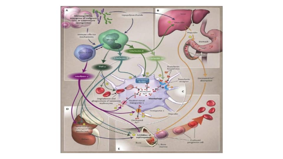
Furthermore, Fiorelli added even greater value by referencing the definitive study in NEJM no less, that explains how all of this ACI chaos happens. I would encourage you to take a moment and read this important article by Weiss & Goodnough, 2005.
Fig. 1 below comes from this key study by Weiss & Goodnough.
So here’s my question: Do you honestly believe that the outrageous complexity that is implied in this picture is accurately or completely captured by the simple, and single misleading blood marker called ferritin?
I would go so far as to suggest that anyone being informed by their healthcare provider that they are “anemic!” should be required to read this 2005 article from NEJM. Then get them to explain how the intracellular protein, ferritin, covers all of those bases and sites of metabolic action. While keeping in mind that our iron-hero, Sir Douglas B. Kell, PhD, world-renowned expert on ferritin, not only thinks, but also has documented that ferritin is found inside the cell, and that the serum level of ferritin (outside of cell) should be zero!
It turns out that this dynamic between iron and the inflammatory process is really important and has been the subject of much research. For those that want to really dig in, please explore the compelling research and writings of Marianne Wessling-Ressnick:
“The evidence that supplemental iron can promote both [emphasis added] infectious and chronic inflammatory disease is clear…”
Has your healthcare practitioner mentioned this known fact about how iron feeds pathogens?
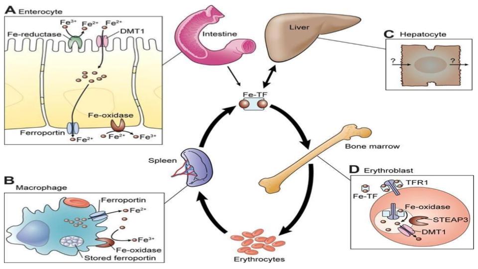
Continuing down this rabbit hole of iron truth are the compelling articles by Nancy Andrews, MD, PhD. She seems to have a knack for simplifying what, at times, can be a most daunting topic. I simply love her diagram of the process of iron recycling, presented as Figure 2, which highlights the dynamic and fluid aspects of iron metabolism.
See how iron is meant to be in constant circulation? The process of iron inside the human body is an elegant square dance where movement and motion are mandatory.
Why are we using an intracellular iron-storage protein, i.e. ferritin, to assess the complexity, importance and elegance of copper<>iron metabolism? Ferritin is not supposed to be in the serum in the first place!
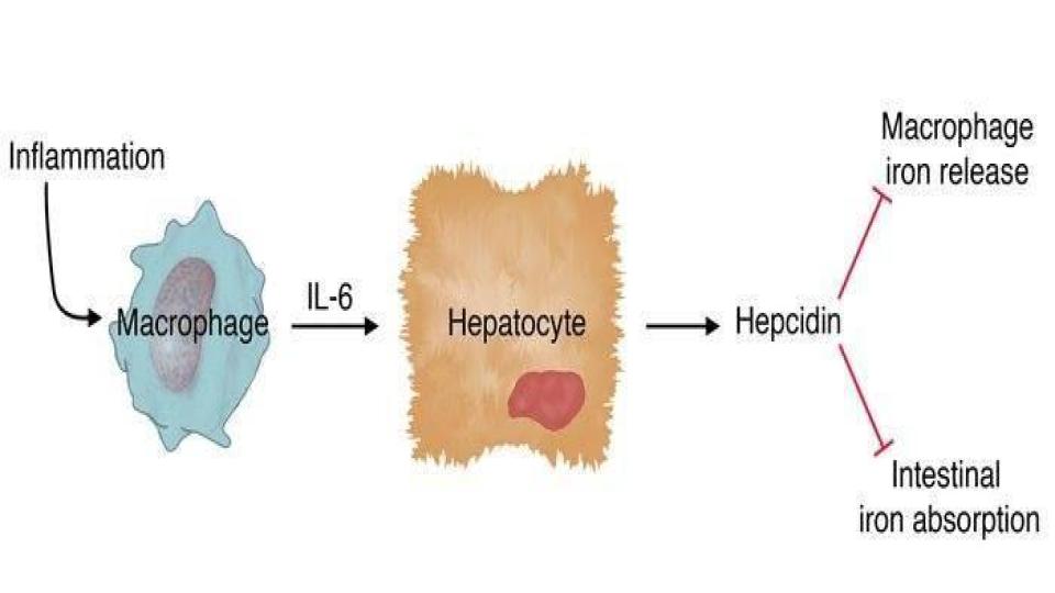
An even better, crisper and shorter article by Dr Andrews is one where she draws the known link between the inflammatory cytokines and their notable impact on the expression of hepcidin that then affects the retention of iron inside the macrophage.
What I especially love about this article is the very simple, but profound diagram of:
How inflammation affects the macrophage,
Which causes the release of the inflammatory cytokine, IL-6,
Which stimulates the synthesis and expression of hepcidin
That then blocks macrophage iron release and blocks intestinal iron absorption.
This is depicted at Figure 3 below.
An important question for those who are still taking iron supplements, yet are experiencing no change in their iron status: Has your doctor explained how iron supplements cause inflammation that then triggers the actions depicted in this diagram?
Here is the link to Dr Andrews’ insights about this cytokine<>hepcidin dynamic:
Andrews, N.C. (2004). “Anemia of Inflammation: the Cytokine-Hepcidin Link.” www.ncbi.nlm.nih.gov/pmc/articles/PMC398435/pdf/JCI0421441.pdf For those that are never satisfied with enough, here are some bonus reads that will shed important light on these iron dynamics, particularly as it relates to the dampening effect that inflammation has on the accessibility of iron in the human body: Karolnek, T., Hamza, I. (2015), “Macrophages and iron trafficking at birth and death of red cells.” www.ncbi.nlm.nih.gov/pmc/articles/PMC4424413/ Nairz, M. et al. (2016). “Iron deficiency or Anemia of Inflammation?” www.ncbi.nlm.nih.gov/pmc/articles/PMC5065583/
Now let’s get down to some brass tacks, did you know that brass is made of copper? What’s really going on here? Where is copper/Cp in all of these diagrams? I thought ceruloplasmin, especially via its ferroxidase function, was critical to these iron issues?
Patience, grasshopper, you are absolutely right! I was just about to point that out!
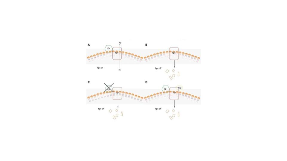
We have discussed this several times before, but the ultra-gifted Italian researcher, Giovanni Musci, has clearly demonstrated how ceruloplasmin is essential for the proper functioning of ferroportin to allow proper and natural iron release from cells, and macrophages, in particular.
This is pointed out in Figure 4 below, please pay particular attention to diagrams A, and note in diagram D how hepcidin presence kills the iron-egress function of ferroportin and also disempowers the supporting role of ceruloplasmin in these critical dynamics to move iron out of the cell.
That is the gift of the ferroxidase function of ceruloplasmin as it ensures proper iron mobilization and circulation in the body. If you haven’t already done so, please enjoy reading this seminal study by Dr Musci:
To me, Figure 3 absolutely sums it up. The only word missing in that simple diagram is the word; iron. It is, indeed, the pivotal agent to cause inflammation.
So, what to do? Here are some suggestions.
Stop thinking that you have a deficiency of iron.
Start thinking that you likely have anemia of chronic inflammation, a condition that is caused by a lack of bioavailable copper, and excess, unbound iron sequestered in the tissues.
Start understanding the complexity and elegance of copper<>iron metabolism
In the event you are told you are “anemic,” do not panic, simply ask the doctor to:
Read the NEJM article by Weiss & Goodnough.
Ask the doctor to explain how the intracellular storage protein, ferritin, is related to and regulates all facets of the iron dynamics shown in that article (our fig 1 above)
Request the full Monty Iron Panel.
Be sure to add a marker for hemoglobin, esp. given that this accounts for 70% of the iron in your body, as opposed to the ~10% that is represented in ferritin.
Please get these additional blood tests properly interpreted before taking any action.
Start the Root Cause Protocol, particularly in the event you have copper<>iron dysregulation
Start donating blood on a regular basis 2 times/year for menstruating women 4-6 times/year for menopausal women or men
Have faith that your body will return to homeostasis, as well as proper metabolic balance.
Start to share these truths & actions with your loved ones.
We are engaged in a wholesale “rewiring” of the mind and our understanding of what factors are genuinely important to assess our copper<>iron metabolism.
These two critical metals that shape the function of the liver are joined at the hip of the ferroxidase enzyme function of ceruloplasmin. It is vitally important that you internalize that and share it with your healthcare practitioner.
I truly hope you find this latest treatise on copper<>iron dynamics helpful and healthful! Have a blessed weekend!
Have I got your attention? I sure hope that you are well rested for this one! This iron toxicity post is, however, a game changer!
I frequently hear from folks who try to tell me that their thyroid Rx meds simply won’t work unless their ferritin is ~100 ng/mL. It is frightening to think there are endocrinologists who believe in this.
I realize that’s a bit harsh, but today I am re-defining the landscape of our understanding about the copper<>iron dynamic.
Let me share a recent conversation that I had with Sir Douglas B. Kell, PhD, knighted in 2014 for his pioneering and penetrating research into ferritin (“Iron Behaving Badly,” 2009a) and whose research lab is at the University of Manchester in Great Britain. It was an honor just to be able to spend some time with him, and also a bit humbling when I realized we were the same age! Dr Kell had actually watched my video, liked it and even said that I was “spot on!” re the pathogens and critters living on that iron.
The highlight of the conversation, however, was when I asked him, point blank, what is the ideal level of ferritin in the serum? With no hesitation, he answered: “zero!” I said, “Excuse me?” Did you really just say, “zero? And he simply smiled! I, then pointed out, “Do you realize that you are in violent opposition with every practitioner on this planet? He merely smiled.
He went on to comment:
“Morley, a ferritin level is not a sign of iron vitality. It is a sign of tissue pathophysiology. The only time ferritin shows up in the serum is when organ cells are breaking down. By the time that ferritin protein shows up in the blood, the iron inside it is likely already released. Note: each molecule of ferritin can hold 4,500 atoms of iron. The ferritin showing up in the blood is most likely the protein, without the iron”
OK, so that short, but engaging conversation absolutely rattled my cage, literally and figuratively. It’s worth noting that ferritin is, in fact, a cage that can hold all those 4,500 iron atoms, as noted above. As we read on, please remember that ferritin comes in two types: Heavy-chain ferritin with ferroxidase function, and Light-chain ferritin with no ferroxidase function.
So that life-changing conversation two weeks ago, really got me to thinking and reflecting on what else I didn’t know about copper<>iron metabolism. It didn’t take the universe too long to drop the bomb in my lap, and that is exactly what occurred this morning.
I decided to do something that I had never done:
Google: Hemosiderin Ferroxidase
I believed I hit the Google jackpot! Up came a blockbuster article:
Please read this article. You can print out a copy for your doctor. Please request that they read it, too! In my humble opinion, it will save your life. Yes, I believe it is that important.
Knowing that there are those that will not take the time to hunt down that article let me quote some of the most compelling passages:
“Rats fed copper-deficient diets [that would be most of us rats, too] had no detectable active [emphasis added] serum ceruloplasmin, which indicates that they were functionally [emphasis added] copper deficient.”
The phrase, copper deficiency, is code for low to no functionality, not lack of the mineral. And this is true of iron, too, I might add but that is topic for another post.
“Animals deficient in copper [i.e. low in ferroxidase function] have an increased amount of hemosiderin.”
Has your doctor spoken to you about hemosiderin levels? Hemosiderin is where iron gets stored in a bank vault with no key. Hemosiderin is an adaptation to the greek word, “siderophore” that means “iron carrier” as depicted in the figure below.
“Additionally, numerous studies have shown that animals fed iron supplemented diets [that would be most of us] have increased amounts of hemosiderin” (Chua-Anusorn et al, 1999; Richter, 1984; Whittaker et al, 1996)
“Therefore, it is possible that any condition which results in iron being loaded into ferritin via its own ferroxidase or by overwhelming the ability of the ferroxidase to load iron into ferritin properly [emphasis added], may result in increased hemosiderin formation.”
The significance of what this team discovered is that when ferritin does not have ferroxidase (as in the light-chain ferritin), the key cellular enzyme outside of ferritin’s own enzyme, the iron does not get loaded into the ferritin properly. It means that the faulty ferritin protein becomes essentially hemosiderin. This is a mind-bender!
Has your healthcare practitioner told you that ferroxidase enzyme is essential for proper iron loading into your ferritin?
“A number of studies suggest that copper plays an integral role in the cellular iron homeostasis, due to the fact that defects in co“We provide data that copper deficient rats [low in ferroxidase function] have an unusual deposition of iron in their hepatocytes (liver cells), indicating the formation of hemosiderin. We also provide data that copper deficiency results in an increased iron content of the hemosiderin fraction isolated from rat liver.”
“Additionally, we demonstrated that these results are associated with a lack of active copper-containing enzyme which has been shown to safely load iron into ferritin, in vivo.”
Copper metabolism, as well as dietary-copper deficiency have been shown to have profound [emphasis added] effects on cellular and systemic iron homeostasis.” (Chen, et al, 2006; Harris & Gitlin, 1996; Lee et al, 1968; Roeser et al, 1970)
“Dietary copper deficiency has been shown to induce an anemia that was not ameliorated by iron supplementation, however, the condition was completely reversed upon copper supplementation.” (Lee et al, 1968; Roeser et al, 1970)
“When iron is loaded into ferritin, in vitro, via its own ferroxidase activity, the ferritin is damaged.” (deSilva et al, 1992; Van Eden & Aust, 2001; Welch et al, 2001, 2002)
“This is intriguing because hemosiderin is thought to be a degradation product of ferritin.” (Miyazaki et al, 2002; O’Connell et al, 1986; O’Connell & Peters, 1987)
I get it; trust me, how hard to grasp the information before us! But this is a key development in our collective efforts to push back the tides of insanity and the lack of integrity of how copper<>iron metabolism are understood and addressed within the world of conventional medical and nutritional treatments. All is not as we’ve been led to believe.
These results and the implications of these results should send a collective chill down all of our spines!
My take on reflecting upon my conversation with Dr. Kell, as well as after reading this stunner of an article:
Ferritin does not belong in the serum, it should only be found inside the cell where it does its work to support the ongoing need for cellular iron proteins and iron-sulfur clusters.
When ferritin does show up in the serum, it is a sign of pathophysiology, not a sign of “iron vitality!” This is a complete reversal of what we have been trained to believe.
Ferritin, which is inside the cell must have access to additional ferroxidase enzyme inside the cell to load iron properly. Has your healthcare practitioner mentioned the need for ferroxidase enzyme that only cellular ceruloplasmin can provide?
When ferritin does not have access to this key ferroxidase enzyme, the iron does not load properly into ferritin, it gets damaged and takes on the properties of hemosiderin, and when that happens, the access to that iron becomes compromised.
Hemosiderin builds up in our tissues and organs as we age. Imagine that! As the “stressors!” of life build, the body’s ability to make ferroxidase drops, and iron builds up in the liver, lungs, spleen, kidneys, lymph nodes, bone marrow & eyes. Those all of the key sights for chronic disease? The only major site missing is the brain but I am confident that it is there, too.
Wow, those findings seem to further validate and support the Root Cause Video.
Are you all beginning to connect these dots?
The iron in hemosiderin is insoluble and allegedly does not come out. I find that hard to believe, but it is the storage equivalent of a bank vault with no key (more on that later).
Most importantly, hemosiderin never shows up in any blood test. It is only found in the tissue of those organs noted above and requires either a needle biopsy or the use of sophisticated and expensive scanning equipment to detect it.
Now, here’s the best part. This is how the article comes to a close:
“However, the mechanism(s) by which copper is involved in iron metabolism remains largely unknown.”
I truly believe that that sentence is a factual statement, but the research knowledge is, indeed, there, it is just that conventional medical education elects not to teach this truth. Therefore, it remains “unknown” as it too, is placed in an educational bank vault!
This is very much in keeping with the classic description of medicine: “Not known, because not looked for”
And the article does add further insights to bring the article to a provocative close with this point.
“It is possible that a copper-containing enzyme, such as ceruloplasmin, may play a role in iron metabolism due to its ferroxidase activity and its ability to load iron into ferritin.” (deSilva & Aust, 1992; Guo et al, 1996; Juan et al, 1997; Juan & Aust, 1998; Reilly et al, 1998; Reilly, 1999; Reilly & Aust, 1998)
My question to you all: Do you think the iron researchers are at all confused about the central role of ferroxidase activity in iron metabolism? I’m mightily impressed, however, by the scope of the research that exists to back up these mineral truths.
Well, we’ve covered a lot of ground today. I know that this post introduces several new concepts and some challenging material, but it also builds on mineral concepts that we’ve been harping on here at the MAG FB Group for the past 18 months.
I will await the comments, the questions and the inevitable pushback. We are forging new ground here. I’m delighted that we have this opportunity to learn together and apply these ground breaking truths in our respective lives and the lives of those we care for the most.
When you or your practitioner suspect that you have any issues with your proper copper<>iron status, these are the many elements that are intimately involved in your copper<>iron metabolism and thus should be the basis of your quest for proper and complete answers. Note the dual nature of many of these elements:
Magnesium RBC
Ferrous (Fe II) iron
Ferric (Fe III) iron
Ferritin Heavy-chain form
Ferritin Light-chain form
Ferritin mitochondrial form which is for the health of brain, heart, liver, thymus, and gonads
Cuprous (Cu I) copper
Cupric (Cu II) copper
Oxygen, found on hemoglobin, inside ferroxidase enzyme and inside ferritins
Ceruloplasmin with ferroxidase (FOX) function
Ceruloplasmin without ferroxidase function and only acute phase reactant response
Anemia of Iron Deficiency (indicates an absolute deficiency of iron)
Anemia of Chronic Inflammation (Indicates an absolute deficiency of bioavailable copper)
If you think just measuring your ferritin status, despite the fact that it exists in three states noted above. Then we have no clue what the blood test is really assessing. Which you would think is a clinically valid and metabolically responsible approach! I encourage you to keep reading.
What I want to stress with you in this post is that anemia does not mean iron deficiency!
I know how outrageously heretical that statement is. But I also know unlike your healthcare practitioner that true and absolute iron deficiency is next to impossible on a planet that has iron comprising 36% of the Earth’s composition, thereby making it the #1 element on the planet.
What is critical to this dynamic is understanding the slight-of-hand that is appearing with increasing frequency in the leading iron research studies.
Iron deficiency means lack of functional iron!
I have now seen it in three different research studies. The iron researchers stating this know that it is bioavailable copper that is key to making iron functional. They know this truth, and by now, I’m hoping you do, too!
I would sincerely ask you to do right now, is take 30 minutes out of your day, and read this vital research on a new discovery in the battle against neurodegeneration:
Yes, this article has big words in it. But this article has some of the most important insights that I have read about iron in a long time which are:
Mitochondrial ferritin (FtMt) is a novel protein localized to the mitochondria.
FtMt is expressed in restricted tissues including the brain, heart, kidney, thymus and gonads.
FtMt lacks iron regulatory elements and its expression is increased by oxidative stress. [That is a big deal!]
FtMt is increased in the cerebral cortex of Alzheimer disease (AD) patients and in the substantia nigra (SN) of individuals with Parkinson’s disease (PD) and Restless Leg Syndrome (RLS).
The presence of FtMt prevents cell death induced by oxidative stress and neurotoxic proteins.
The two most important sentences of this study, in my humble opinion, are:
“…FtMt, which also possesses ferroxidase activity.”
Which means this critical protein; ferroxidase (FOX) can’t do anything without bioavailable copper, aka ceruloplasmin (Cp = FOX).
“Mitochondrial Ferritin (FtMt) overexpression showed a neuroprotective effect against hydrogen peroxide (H2O2) induced oxidative stress and amyloid beta-induced neurotoxicity in neuroblastoma cells”
Bioavailable copper coming to the rescue of tissue being assaulted by iron-induced oxidative stress!
What this means is that a healthy body has a naturally occurring mechanism to metabolize neurotoxic iron that is only possible with mitochondrial ferritin that depends on ferroxidase function. This is only possible in a body that has optimal levels of bioavailable copper.
Here is my take:
How much does your doctor really know about iron toxicity?
There is no such thing as iron metabolism as it is only copper<>iron metabolism.
Bioavailable copper regulates iron status and lack of copper creates iron toxicity.
You are not anemic from iron deficiency but lack of functional iron, which causes anemia of chronic inflammation.
Iron supplements and iron infusions make this dynamic worse and may even develop neurodegeneration.
Please learn and understand your true mineral status via HTMA and blood testing.
Please get a “Copernican (RCP) inspired” consult to learn how the stress mosaic of your life created the mineral dysregulation that you are now expressing. https://therootcauseprotocol.com/rcpc-directory/
Please understand that mineral dysregulation created the “Ferrous Wheel” and the consequence creation of symptoms that are expressions from iron-induced oxidative stress.
Please start with the Root Cause Protocol (RCP) – even before you have a consult so you can begin to initiate mineral balance that will then allow metabolic recovery and optimal function. https://therootcauseprotocol.com/about/
“The two most Important Days of your Life: the day you were born, and the day you discover why!” — Mark Twain
I love that quote from Mr Clemmons! He was such a sage. You know he is right; the day you finally figure out why you’re here is magical. It is indelibly etched in my psyche and relates entirely to stepping onto this virtual podium or some would say, bully pulpit! Nevertheless, I’m having the time of my life and I never regret this 2nd birthday.
I also enjoyed the appearance of our light being Jack Kruse, MD. He never fails to stimulate our little grey cells. In any event, I elected to take the dive with the blog on how red light helps to address an underactive thyroid. It was provocative and thoughtful, but do people have any idea where red light comes from inside of our cells?
Red light is generated when there is proper function of cytochrome c oxidase (Complex IV) of the mitochondrial electron transport chain (ETC). Why is that?
Complex IV does not work without bioavailable copper. It is also worth noting that copper and red light have a very symbiotic relationship.
So this blog article is an important clue about what is missing from a healthy thyroid is bioavailable copper. Where is that copper absence greatest? The key enzyme thyroid peroxidase (TPO) works best when there is wholefood vitamin C in the system. Whole food vitamin C is a source of bioavailable copper with tyrosinase at the core. Tyrosinase has a connection to the tyrosines that are being loaded with iodine. I am surprised that I seem to be the only one that sees this obvious deflection of reality by the world of convention obsessed with taking our thyroids hostage.
In any event, that got me thinking and searching this morning for some additional research re this red light phenomenon. What I came across is an absolute blockbuster:
I know you all have very active, demanding and stress-filled lives. But please, take the 30 min it will take you to read through this very important and timely study.
What’s important to understand at the outset that Photobiomodulation (PBM) is considered a cutting edge mechanism for addressing metabolic shortages and dysfunction via the use of low level laser therapy (LLLT). Yes, there’s great buzz about using lasers, LEDs, and related technology. It clearly works to relieve symptoms, but is it just another expensive crutch or just more diversion from the real mineral and metabolic issues? I would question whether fundamentally, does PBM correct the underlying issues that are causing this metabolic chaos?
I could spend the rest of this month commenting on what I learned reading this critical article, but what i want to stress are just a few key points:
Rapidly growing cells prefer cell death instead of fixing the damaged mitochondria
Electromagnetic light must ionize biological systems that it effects
Irradiation [by] external electromagnetic energy can increase and substitute [emphasis added] for endogenous ATP production
Radiation of red light (at 628 nm on the Light Spectrum) results in expression shifts of 111 genes in Hs27 human fibroblasts (Zhang, 2003). I would encourage you to read that sentence again! Zhang, Z., Song, S., et al. (2003). “cDNA MicroarrayAnalysis of Gene Expression Pro¢les in Human Fibroblast Cells Irradiated with Red Light.” https://pubmed.ncbi.nlm.nih.gov/12713592/
These 111 expressive shifts were grouped into 10 functional categories that promoted extracellular matrix proteins, orchestrated an increase in adenosine triphosphate (ATP) production, and also orchestrated an increase in cellular stress resistance [again, emphasis added]. That is the granddaddy of them all.
Retrograde signaling [a form of mitochondrial stress response] can be mediated [another way of saying stimulated] by:
Mitochondrial membrane potential (caused by copper<>iron dysregulation)
Reactive oxygen species (ROS) (caused by copper<>iron dysregulation)
Changes in calcium flow (caused by iron dysregulation affecting magnesium status)
Nitric oxide binding to cytochrome c oxidase, again, caused by iron dysregulation which is spelled out here:
To those wishing to dig in even deeper, please follow this trail. These are both blockbuster articles, as well: Lane, N. (2006). “Power Games.” https://www.nature.com/articles/443901a
Do you follow my point? There is clearly mitochondrial stress and what we are now learning is that it reaches over and touches the nucleus. (Please see the Fig. 1 below)
Think this iron-induced oxidative stress, as noted in points 6a thru 6d might just be the true source of the genetic defects that we’re supposed to believe come from mars?
These exogenous electromagnetic machines emit a red light that clearly helps to correct this chaos and confusion inside our cells. So, why am I not excited at this prospect?
You were not born with a red light deficiency. You were born with excess, unbound iron along with a notable shortage of bioavailable copper. I believe that is a fact in the research.
I know that these powerful and penetrating research studies are lighting the way for what the true issues are; just as we’ve been saying for four years.
Please don’t misunderstand my comments above; I am very excited with these developments. In fact, I’m seriously contemplating adding red light to the Root Cause Protocol. It’s that powerful, but I do so as a means to an end (crutch). The ultimate purpose in our efforts on Magnesium Advocacy Group (MAG) is to create more ceruloplasmin and allow the body to regain its mineral and metabolic homeostasis.
I was also fascinated to learn that the great Otto Warburg knew much of this back in 1931 when he received the Nobel Prize for his work on none other than the mitochondrial respiratory enzyme that was related to hemoglobin, which turns out to be cytochrome c oxidase.
We have learned a thing or two since 1931, but he was on the trail. Here is what Otto Warburg had to say about his Nobel speech award at the banquet in December of 1931:
What Dr Warburg did know was that cancer cells had three key conditions: 1) Low pH 2) Low oxygen and 3) Low ATP.
Intriguing! That means when oxygen is low, the cell is forced to produce ATP via fermentation, and not via oxygen, which is its preferred route.
It turns out that Otto Warburg was studying the key mitochondrial enzyme (Cytochrome c Oxidase) that is fuelled by bioavailable Copper and he theorized the very three cellular conditions that are caused by excess, unbound iron.
Absolutely fascinating! Forgive me, but my mind’s blown at this point!
Now here’s what you may not know: When the mitochondria have access to oxygen to “burn its food-based fuel,” it is relying on copper to oxidize the sugars and fats to enable the production of ATP.
Here is the breakdown:
1 Unit of fat yields 120 ATPs
1 Unit of sugar yields 32-34 ATPs
When the cell is forced to go anaerobic (because of a loss of copper and/or excess of iron) and fermentation is used to breakdown the sugars, the yield is 2 ATPs (Please note the prevalence of magnesium (Mg) ions needed to make the anaerobic citric acid cycle work properly)
The methodical depletion of copper in our diet over the last 100yrs, and the known process of iron fortification that have introduced absurd levels of iron into our bodies have lowered our ability to use “gas” that can do 120 MPG or 32 MPG. We are forced to ferment our foods and burn up whatever magnesium we might possibly have. For those of you wondering how and why you have chronic fatigue please look no further.
Just know that we are on the right track with our focus on MAG and for those that are just joining us, please take the time to familiarize yourself with the Video and the Root Cause Protocol.
Taking the time to do that just might save your life, and it will certainly save you years of misery groping for a solution in a conventional medical system that is only committed to treating your condition. The latest gadget in their arsenal of goodies is their laser that helps, but only treats your condition.
MAG is fully committed to allowing you to regain your mineral and metabolic balance – naturally.